
2/16/2014 ED Ultrasound for Undifferentiated Hypotension Nate Teismann MD Director of Emergency Ultrasound Emergency Ultrasound Fellowship Director Dept of Emergency Medicine, UCSF nathan.teismann@ucsf.edu 1
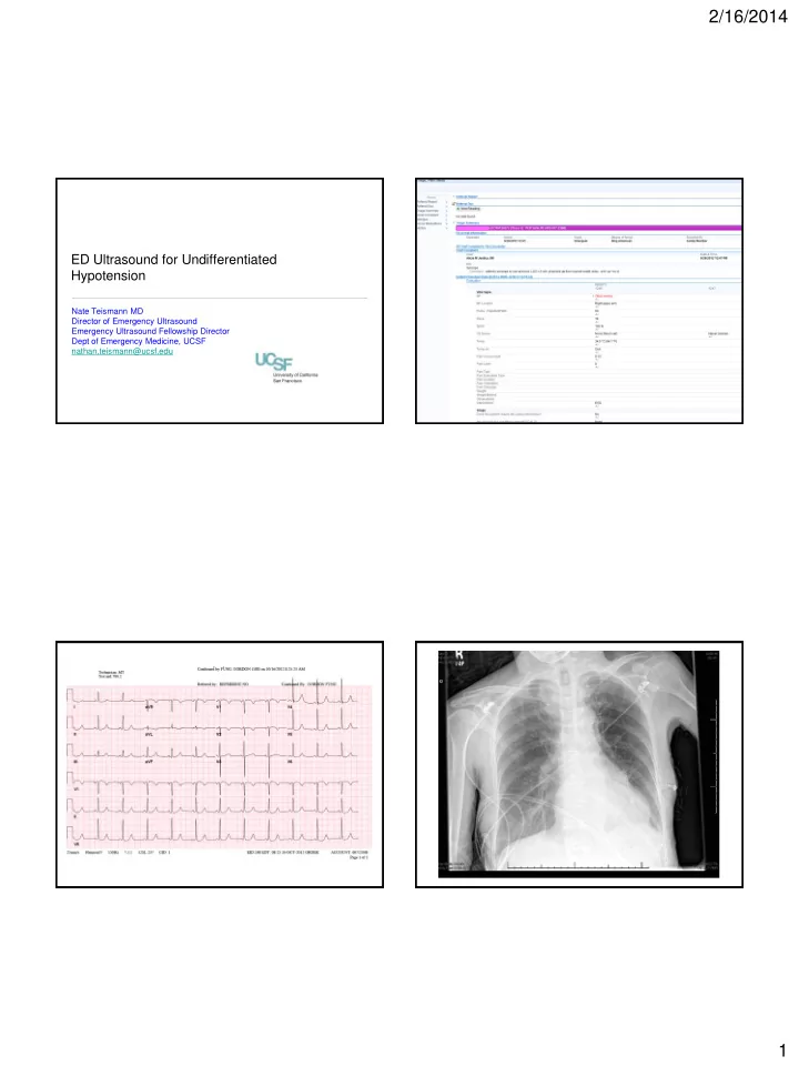
2/16/2014 70 F “not feeling well” + syncope Why is this patient sick? Your approach with US: • Heart: • PMH: Unknown • Is there an effusion/tamponade? • Meds: Unknown • What is the LV doing? • Pale, Diaphoretic • Is the RV under strain? • Afebrile, Faint crackles, • IVC: tachypneic, otherwise • What is the CVP? unremarkable exam • Lungs, Deep LE Veins, Aorta: BP = 62/38 • Is there a pneumothorax, pulmonary edema, or lower lobe PNA? • Is there a DVT or Aortic Pathology? Can’t I just try a fluid challenge? • Fluid therapy is mainstay of treatment...except when it’s not. • Other interventions: 80% sepsis or hypovolemia • Pressors/Inotropes? • ED procedures (pericardiocentesis, thoracostomy)? • To OR? 2
2/16/2014 QuickTime™ and a VC Coding QuickTime™ and a H.264 decompressor are needed to see this picture. Step 1: What is the heart doing? • Is there an effusion present? QuickTime™ and a VC Coding • If so, are there signs of tamponade? • Clinical > US findings! QuickTime™ and a QuickTime™ and a VC Coding VC Coding 3
2/16/2014 QuickTime™ and a VC Coding QuickTime™ and a VC Coding Step 1: What is the heart doing? QuickTime™ and a VC Coding • Is there an effusion present? • If so, are there signs of tamponade? • LV size and systolic function • Normal, Poor, Hyperdynamic • EPSS QuickTime™ and a VC Coding 4
2/16/2014 Mitral E-Point Septal Separation QuickTime™ and a QuickTime™ and a QuickTime™ and a VC Coding VC Coding VC Coding EPSS > 1cm predicts EF < 50% QuickTime™ and a DV - NTSC decompressor are needed to see this picture. Step 1: What is the heart doing? Step 1: What is the heart doing? • Is there an effusion present? • Is there an effusion present? • If so, are there signs of tamponade? • If so, are there signs of tamponade? • LV size and systolic function • LV size and systolic function • Normal, Poor, Hyperdynamic • Normal, Poor, Hyperdynamic • EPSS • EPSS • RV size and function 5
2/16/2014 QuickTime™ and a H.264 decompressor QuickTime™ and a are needed to see this picture. H.264 decompressor are needed to see this picture. QuickTime™ and a H.264 decompressor are needed to see this picture. Size: Normal RV < 70% LV Signs of acutely elevated R-sided pressures: • 1) RV enlargement • 2) RV hypokinesis (possibly with apical sparing = McConnell’s) • 3) Flattening of the interventricular septum (D-shaped LV) RV LV • 4) RA and IVC dilation QuickTime™ and a JVT/AVC Coding decompressor are needed to see this picture. LV RV QuickTime™ and a H.264 decompressor are needed to see this picture. 6
2/16/2014 QuickTime™ and a QuickTime™ and a VC Coding H.264 decompressor are needed to see this picture. Acute! QuickTime™ and a JVT/AVC Coding decompressor are needed to see this picture. Chronic QuickTime™ and a QuickTime™ and a VC Coding H.264 decompressor are needed to see this picture. 1) Mild-Mod RV Enlargement 1) Severe RV Enlargement 2) No RVH 2) RVH 3) McConnell’s Yes 3) McConnell’s No Thrombus “In - Transit” Step 1: What is the heart doing? Step 2: What is the CVP? • Is there an effusion present? • Look at IVC: 2 parameters • If so, are there signs of tamponade? • 1) Maximum diameter • LV size and systolic function • 2) Caval Index (% collapsibility with inspiration) • Normal, Poor, Hyperdynamic • EPSS • RV size and function • Signs of elevated R-sided pressures? • VERY large RV with RVH suggests chronic pulmonary HTN 7
2/16/2014 CVP High Step 2: What is the CVP? QuickTime™ and a VC Coding • Look at IVC: 2 parameters • 1) Maximum diameter at hepatic vein inlet • > 2cm suggests CVP >10 QuickTime™ and a Sorenson Video 3 decompressor are needed to see this picture. • 2) Caval Index (% collapsibility with inspiration) CVP Low • > 50% collapse with passive inspiration predicts CVP < 8 QuickTime™ and a Normal VC Coding QuickTime™ and a QuickTime™ and a VC Coding VC Coding Normal CVP Low 8
2/16/2014 Step 2: What is the CVP? • Look at IVC: 2 parameters QuickTime™ and a VC Coding • 1) Maximum diameter • > 2cm suggests CVP >10 • 2) Caval Index (% collapsibility with inspiration) • > 50% collapse with passive inspiration predicts CVP < 8 QuickTime™ and a VC Coding • Big, dilated, noncollapsible IVC = CVP high CVP High • Small, thin, highly collapsible IVC = CVP low Step 3a: Anything in the lungs? Step 3b: Anything elsewhere? • Is there a blood in the abdomen? • Is there a pneumothorax? QuickTime™ and a decompressor are needed to see this picture. • Is there a AAA? • Is there a consolidation? • Are there a DVT? • Are there B lines? QuickTime™ and a VC Coding QuickTime™ and a QuickTime™ and a VC Coding VC Coding QuickTime™ and a AVC Coding decompressor are needed to see this picture. 9
2/16/2014 QuickTime™ and a decompressor are needed to see this picture. QuickTime™ and a VC Coding Review QuickTime™ and a VC Coding QuickTime™ and a QuickTime™ and a Motion JPEG OpenDML decompressor VC Coding are needed to see this picture. QuickTime™ and a VC Coding PNA, Sepsis, CHF, cardiogenic shock hypovolemia 10
2/16/2014 QuickTime™ and a V C Coding QuickTime™ and a QuickTime™ and a V C Coding QuickTime™ and a decompressor H.264 decompressor are needed to see this picture. are needed to see this picture. QuickTime™ and a V C Coding Pericardial Effusion with QuickTime™ and a V C Coding Impending Tamponade QuickTime™ and a V C Coding Acute Massive PE Thanks! 11
Recommend
More recommend