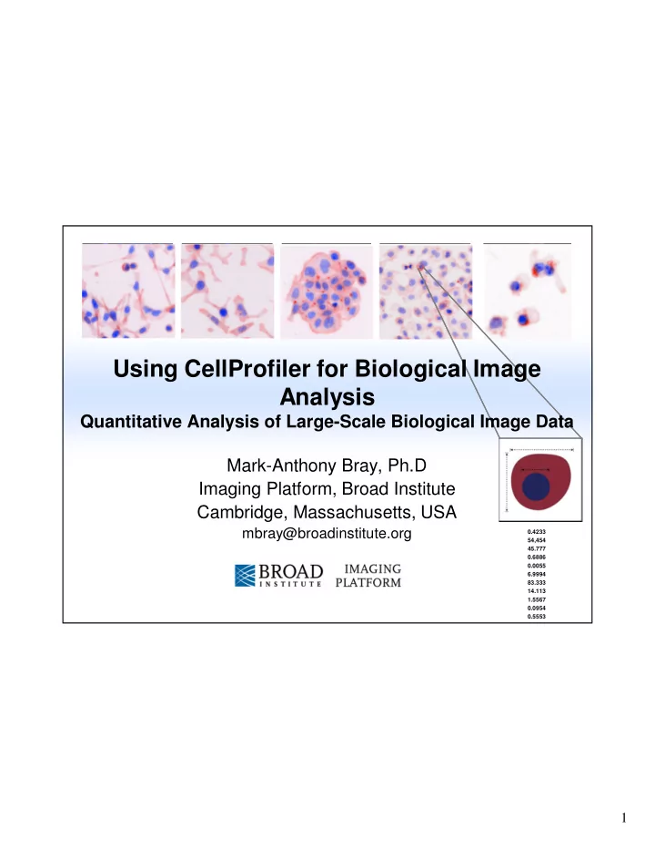

Using CellProfiler for Biological Image Analysis Quantitative Analysis of Large-Scale Biological Image Data Mark-Anthony Bray, Ph.D Imaging Platform, Broad Institute Cambridge, Massachusetts, USA mbray@broadinstitute.org 0.4233 54,454 45.777 0.6886 0.0055 6.9994 83.333 14.113 1.5567 0.0954 0.5553 1
Summary • Background on image-based screening • Introduction to CellProfiler considerations in image analysis • Construction and use of a pipeline for analyzing typical image data • Measurement export and preparation for additional analysis 2 2
Images Contain A Wealth Of Information http://www.microscopyu.com Image: Javier Irazoqui 3 3
Visual Appearance Indicates Biological State • Images contain a wealth of Localization biological information • That information can be quantified mRNA or protein levels • Automatic image analysis is – Objective – Quantitative, with statistics morphology – Can measure multiple properties at once for every cell – Distinguishes subtle changes, even those undetectable by eye … + hundreds of other features – Faster, less tedious 4 4
High-Content Screening Cells or organisms in multiwell plates, each well treated with a gene or chemical perturbant Cell measurements Automated microscopy (size, shape, (any manufacturer) intensity, texture, etc.) Data exploration & machine learning Ray Anne Jones Carpenter 5 5
Software Overview Image Analysis & Quantification Image-centric Data Analysis • Available from www.cellprofiler.org • Free, open source (Python) • Software available for Windows, Mac and Linux 6 6
CellProfiler: Overview • Process large sets of images • Identifies and measures objects • Export data for further analysis • Goal: Provide powerful image analysis methods with a user-friendly interface • Philosophy: Measure everything, ask questions later... • Support data analysis based on individual cells 7 7
Typical CellProfiler Pipeline Workflow • For image-based assays, the basic objective is always to – Identify cells/organisms – Measure feature(s) of interest • The uniqueness of each assay comes in – Deciding what compartments to identify and how to identify them – Determining which measure(s) are most useful to identify interesting samples 8 8
Typical CellProfiler Pipeline Workflow 9 9
The CellProfiler Interface Module help Add or remove modules Change module position • Pipeline panel: Displays modules in pipeline – Modules executed in order from top to bottom 10 10
The CellProfiler Interface Load pipeline by double-clicking on it View images by double-clicking on the filename • File panel: Displays files in default image folder 11 11
The CellProfiler Interface • The figure window has additional menu options • Toolbar menu: Pan, zoom in/out • CellProfiler Image Tools – Image Tool (also displayed by clicking on image) – Interactive zoom – Show pixel data (location, intensity) 12 12
The CellProfiler Interface Input folder: Contains images to be analyzed Output folder: Contains the output file plus exported data and images • Folder panel: Change default input and output directories – Usually these should be separate folders 13 13
The CellProfiler Interface • Settings panel: View and change settings for each module – Clicking on a different module updates the settings view 14 14
Module Categories • File processing: Image input, file output • Image processing: Often used for pre-processing prior to object identification • Object processing: Identification, modification of objects of interest • Measurement: Collection of measurements from objects of interest • Data Tools: Measurement exploration, measurement output 15 15
̶ The First Module: LoadImages • Loads an image set A group of related images to be processed DNA GFP • Related how? Depending on the imaging device, one file may represent – One channel at one imaging location – Multiple channels at one imaging location – Multiple channels at multiple locations – Etc… 16 16
The First Module: LoadImages • Can use text matching to define the difference between images in a set All images stained for GFP have the text Channel1- in the name Assign each image a meaningful name for downstream reference Same for DNA images ( Channel2- ) 17 17
Object Identification • Once the images are loaded, how do you find objects of interest? • Step 1: Distinguish the foreground from the background by picking a good threshold • Step 2: Identify objects as regions brighter than the threshold • Step 3: Cut and join objects to “improve” their shape 18 18
Primary Object Identification • Many options for thresholding, cut and join methods, etc. 19 19
Thresholding • Definition: Division of the image into background and foreground Frequency • What is the best threshold value for dividing the intensity into foreground and background pixels? Pixel values • Method: Pick the method that provides the best results – Otsu: Default - Good for readily identifiable foreground / background – Background, RobustBackground: Good for images in which most of the image is comprised of background 20 20
Thresholding • Correction factor – Multiplication factor applied to threshold – Adjusts threshold stringency/leniency – Setting this factor is empirical • Upper/lower bounds – Set safety limits on automatic threshold to guards against false positives – Helpful for unexpected images: Empty wells, images with dramatic artifacts, etc 21 21
Object Separation • Once the foreground objects have been identified, what next? • • • • • • • • • We need to distinguish multiple objects contained in the same “clump” Images from Carolina Wahlby 22 22
Object Separation Adjust settings to “de-clump” objects • Two step process in “de-clumping” 1. Identification of the objects in a clump 2. Drawing boundaries between the clumped objects 23 23
Object Separation • Clump identification: Two options Peaks – Intensity: Works best if objects are brighter at • center, dimmer at • • • edges 1 2 • • • • 1 Indentations – Shape: Works best if objects have indentations where clumps touch (esp. if 1 2 objects are round) 24 24
Object Separation • Drawing boundaries: Two options – Distance: Draws boundary lines • midway between • • • object centers • • • • 1 – Intensity: Draws boundary lines at dimmest line between objects • Test Mode allows users to view results of all setting combinations 25 25
Object Separation • Additional separation settings: Adjust these settings if objects are being incorrectly split into pieces or merged together Original image Smoothing filter Smoothing filter size = 4 size = 8 • Smoothing: Increase to reduce intensity irregularities which produce over-segmentation of objects 26 26
Object Separation Maxima Original image Maxima Maxima distance = 4 distance = 8 • Suppress Local Maxima – Smallest distance allowed between object intensity peaks to be considered one object rather than a clump – Decrease to reduce improper merging of objects in clumps 27 27
Object Separation • However…. Original image Smoothing filter Smoothing filter size = 4 size = 8 • Adjusting can produce more improper segmentation than it solves • The proper settings are usually a matter of trial and error – The automatic settings are a good starting point, though 28 28
Filtering Invalid Objects Discard objects that fail size criterion or touch the image border • See FilterObjects module for more advanced filtering options 29 29
Primary Object Identification • Segmented objects are colored – Shows if each object has been identified and separated properly • Outlines: Valid objects – Green: Valid – Yellow : Invalid – Touching border – Red: Invalid – Size criterion • Also outputs object count 30 30
Secondary Object Identification • Goal: Identify cell boundaries by “growing” primary objects – Nuclei typically more uniform in shape, more easily separated than cells Approach: Segment nuclei → Seeds for cell segmentation by using a • cell stain channel 31 31
Secondary Object Identification • Methods – Distance-N: Ignores image information • Useful in cases where no cell stain is present – Watershed, propagate, Distance-B: Uses image Distance-N information • Finds dividing lines between objects and background / neighbors • Test mode allows user to view results of all methods Propagation 32 32
Tertiary Object Identification • Goal: Identify tertiary objects by removing the primary objects from secondary objects – “Subtract” the nuclei objects from cell objects to obtain cytoplasm ═ Cells Nuclei Cytoplasm — 33 33
Recommend
More recommend