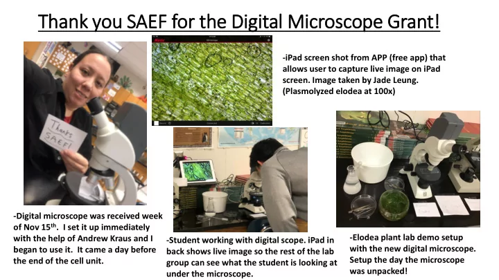

Thank you SAEF for the Dig igit ital Mic icroscope Grant! -iPad screen shot from APP (free app) that allows user to capture live image on iPad screen. Image taken by Jade Leung. (Plasmolyzed elodea at 100x) -Digital microscope was received week of Nov 15 th . I set it up immediately -Elodea plant lab demo setup with the help of Andrew Kraus and I -Student working with digital scope. iPad in with the new digital microscope. began to use it. It came a day before back shows live image so the rest of the lab Setup the day the microscope the end of the cell unit. group can see what the student is looking at was unpacked! under the microscope.
Annotations and scale bars can be directly added to Microscope Im Image Quality images with the user friendly iPad software! iPad image of Elodea Cells at 100x Image with microscope from grant of Elodea Cells at 100x -very hard to distinguish cells & details -clear, crisp image with the ability to see cell details
Classroom use of Microscope & Software Andrew and I tested the software out on our Students working iPads before I with the new presented the new microscope and technology to the digital technology students. software Students Advantages: downloading the Motic camera -Students can open their iPads and see the image I have app on their under the microscope iPads -Absent students can catch up on missed labs -Students can take their own images using their iPads and upload those in to lab reports or on to Google Classroom
Microscope Application in Biology The microscope ($458.10 from Carolina science supply company) was ordered immediately in August after I received confirmation of receiving the SAEF grant. The microscope arrived in November after the cell unit (unit with major microscope applications) but was used throughout the year in many units. Cell Cycle Phases of Photosynthesis Elodea Mitosis Onion Root Plasmolysis Lab Tip Lab Rootbeer Yeast Fermentation Lab Strawberry DNA Extraction Lab Spinach Leaf Tape Impression Guard Cell Lab
Ecology Unit Examples (M (May 2019-added aft fter orig rigin inal l pre resentation date e of f Marc rch/April) l) Students are looking at North Park Lake water to As part of the pre-lab intro I showed students examples of things identify protists as part of the aquatic ecosystem they would find- phytoplankton & zooplankton. Students could see ecology lab. Students took pictures & videos of real-time images of the samples. Images were also taken & labeled their samples. for students who were absent to complete the make up lab.
North Park La Lake 40x -Example of f Pic icture wit ith Dig igital Camera Freshwater macroinvertebrate
Water Bear Ecology Lab: : Pre-lab In Intro Clear, crisp images are easily obtained from the Powerpoint was prepared to help digital microscope and can students understand what they be shared with students. were trying to find in the lab! I Live-footage from the could also show them real-time microscope can be viewed examples of the water bear if they by students on their iPads! had trouble finding one in their sample
Thank you SAEF for the Dig igit ital Mic icroscope Grant! Example of image taken by 9 th grade students. Jade Leung’s Honors Biology & Biology 9 th and 10 th grade students used the microscope throughout the year. Microscope was also shared and made available to other high school science teachers.
Recommend
More recommend