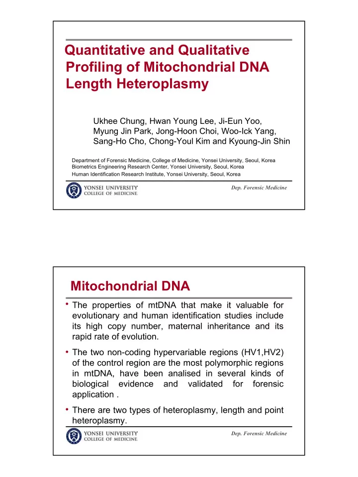

Quantitative and Qualitative Profiling of Mitochondrial DNA Length Heteroplasmy Ukhee Chung, Hwan Young Lee, Ji-Eun Yoo, Myung Jin Park, Jong-Hoon Choi, Woo-Ick Yang, Sang-Ho Cho, Chong-Youl Kim and Kyoung-Jin Shin Department of Forensic Medicine, College of Medicine, Yonsei University, Seoul, Korea Biometrics Engineering Research Center, Yonsei University, Seoul, Korea Human Identification Research Institute, Yonsei University, Seoul, Korea Dep. Forensic Medicine Mitochondrial DNA The properties of mtDNA that make it valuable for evolutionary and human identification studies include its high copy number, maternal inheritance and its rapid rate of evolution. The two non-coding hypervariable regions (HV1,HV2) of the control region are the most polymorphic regions in mtDNA, have been analised in several kinds of biological evidence and validated for forensic application . There are two types of heteroplasmy, length and point heteroplasmy. Dep. Forensic Medicine 1
Subject of Investigation The genetic characteristics of length heteroplasmy have been the subject of the investigation in mtDNA. No guiding criteria for the interpretation of mtDNA length heteroplasmy have been established due to sequencing method limitation. Therefore, In an attempt to investigate mtDNA length heteroplasmy, it is prerequisite to develop a new method capable of complementing sequencing analysis. Dep. Forensic Medicine Samples One hundred unrelated Korean DNAs were extracted from buccal swabs using QIAamp DNA Mini Kit. The two hypervariable regions of mitochondrial DNA were amplified in a PCR mixture of total volume 10.0ul containing 0.05~0.1ng of DNA template. Dep. Forensic Medicine 2
Amplification Primer Set L16144 5’-TGA CCA CCT GTA GTA CAT AA H16410 * 5’- FAM -GAG GAT GGT GGT CAA GGG AC L155 5’-TAT TTA TCG CAC CTA CGT TC H389 * 5’- HEX -CTG GTT AGG CTG GTG TTA GG Theraml Cycling 95 ° C for 11min Initial denaturation 94 ° C for 1min Denaturation 56 ° C for 1min Annealing X 25 cycles 72 ° C for 1min Extension 60 ° C for 45min Final extension Dep. Forensic Medicine Size-Based Separation The PCR products were separated by capillary electrophoresis using an ABI PRISM 310 genetic analyzer (Applied Biosystems). POP 6 was utilized to the resolution of the separation and GS STR A module was adapted as a run module. The resulting data were analyzed using GeneScan software 3.1 (Applied Biosystems) and none of smooth option as an analysis parameter. Dep. Forensic Medicine 3
Confirmation of Length Heteroplasmy To analyze sequence encompassing the polymorphic tracts in the HV1 and HV2 regions, PCR products were using a BigDye Terminator Cycle Sequencing v2.0 Ready Reaction Kit (Applied Biosystems). In order to identify each mtDNA length variant within the heteroplasmic mtDNA mixture, cloning and sequencing were carried out using pGEM-T Easy Vector (Promega). Dep. Forensic Medicine Proportion of Length Variation HV1 HV2 Length Length Length Length Proportions Proportions homo- hetero- homo- hetero- of length of length Size a Size a) variants plasmy plasmy variants plasmy plasmy (bp) (bp) 234 2 0 2 266 2 1 1 235 3 3 0 267 77 63 14 236 34 28 6 268 14 0 14 237 41 0 41 269 7 0 7 238 19 0 19 239 1 0 1 Sum 100 64 36 Sum 100 31 69 The study demonstrated 36 % and 69 % of Koreans show length heteroplamy in the HV1 and HV2 regions. Dep. Forensic Medicine 4
The Peak Pattern of HV1 AAAA C10 ATGC AAAA C9 ATGC AAAA C11 ATGC According to the GeneScan electropherograms, all heteroplasmic mtDNAs were classified into 5 major peak patterns . Dep. Forensic Medicine Sequences of HV1 Sequences A B C D E F Sum AAAA CCCCCTCCCC ATGC 50 50 AAAA CCC T C T CCCC ATGC 3 3 Sequences A B C D E F Sum AAAA T CCCC T CCCC ATGC 3 3 2 2 AAAA CCCCC C CCCC ATGC AAAA C T CCC - CCCC ATGC 1 1 1 1 AAAA CCCCC C CCCC G TGC AAAA C T CCC T CCCC ATGC 1 1 AAAA CCCCC C CCCC C ATGC 6 1 7 AAAA CCC T C C CCCC ATGC 3 3 1 1 AAA C CCCCC C CCCC ATGC AAAA CCCCC T CC T C ATGC 2 2 AAA C CCCCC C CCCC CC GC 1 1 AAA G CCCCC T CC T C ATGC 1 1 AAA C CCCCC C CCCC C ATGC 13 1 14 Sum 64 0 0 0 0 0 64 AA CC CCCCC C CCCC ATGC 8 8 AA CC CCCCC C CCCC CC GC 1 1 AA TC CCCCC - CCCC ATGC 1 1 Sum 0 15 9 8 3 1 36 Dep. Forensic Medicine 5
The mtDNA Peak Patterns of HV1 Type N % a) The most prevalent length variant N % b) A 64 _ 63 98.4 16185T, 16189d 1 1.6 B 15 41.7 16189C 2 13.3 16189C, 16193.1C 13 86.7 C 9 25.0 16189C 9 100.0 D 8 22.2 16189C 1 12.5 16189C, 16193.1C 1 12.5 16189C, 16193.1C, 16193.2C 6 75.0 E 3 8.3 16189C, 16193d 1 33.3 16189C 2 66.7 F 1 2.8 16189C, 16193.1C, 16193.2C 1 100.0 Sum 100 100 a) Proportions of the total number of heteroplasic mtDNA b) Proportions of the most prevalent length variants of each peak pattern Dep. Forensic Medicine The Peak Pattern of HV2 According to the GeneScan electropherograms, all heteroplasmic mtDNAs were classified into 7 major peak patterns . Dep. Forensic Medicine 6
Sequences of HV2 Sequences A B C D F G H I Sum AA CCCCCCCTCCCCCC GC 31 1 1 2 35 AACCCCCCC C T CCCCCCGC 15 12 16 43 AACCCCCC CC T CCCCCCGC 11 8 19 AACCCCCC CCC T CCCCCCGC 1 1 AACCCCCCC - CCCCC - GC 1 1 2 Sum 31 27 21 16 2 1 1 1 100 Dep. Forensic Medicine The mtDNA peak patterns of HV2 Type N % a) The most prevalent length variant N % b) A 31 315.1C c) 28 90.3 249d, 315.1C 3 9.7 B 27 39.1 249d, 309.1C, 315.1C 1 3.7 315.1C 1 3.7 309.1C, 315.1C 14 51.9 309.1C, 309.2C, 315.1C 11 40.7 C 21 30.4 315.1C 1 4.8 309.1C, 315.1C 12 57.1 309.1C, 309.2C, 315.1C 8 38.1 D 16 23.2 249d, 309.1C, 315.1C 1 6.2 309.1C, 315.1C 15 93.8 E 2 2.9 315.1C 2 100.0 F 1 1.4 309.1C, 309.2C, 309.3C, 315.1C 1 100.0 G 1 1.4 310d 1 100.0 H 1 1.4 310d 1 100.0 Sum 100 100 a) Proportions of the total number of heteroplasic mtDNA b) Proportions of the most prevalent length variants of each peak pattern 7
Electropherogram of HV2 The HV2 heteroplasmic peak patterns in GeneScan analysis were very similar to multiple T peaks shown in the middle of homopolymeric C-stretch in sequencing electropherograms. Dep. Forensic Medicine Conclusions We established a new strategy for profiling length heteroplasmies. Classification of mtDNAs into several types of peak patterns is believed to offer a useful means of determining genetic identity by increasing mitochondrial DNA haplotype diversity. The developed method will present a promising tool for the diagnosis of several common diseases which are etiologically or prognostically associated with mtDNA polymorphisms. Dep. Forensic Medicine 8
Recommend
More recommend