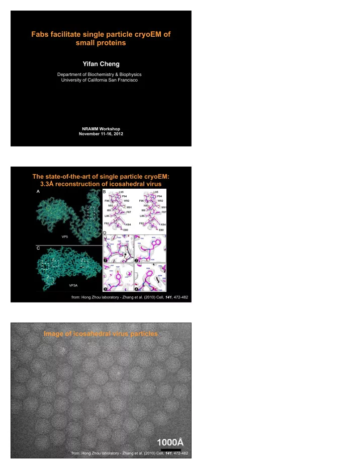

Fabs facilitate single particle cryoEM of small proteins Yifan Cheng Department of Biochemistry & Biophysics University of California San Francisco NRAMM Workshop November 11-16, 2012 The state-of-the-art of single particle cryoEM: 3.3Å reconstruction of icosahedral virus from: Hong Zhou laboratory - Zhang et al. (2010) Cell, 141 , 472-482 Image of icosahedral virus particles from: Hong Zhou laboratory - Zhang et al. (2010) Cell, 141 , 472-482
How to apply single particle cryoEM to study small proteins? * Images of individual small molecule do not contain sufficient structural information for accurate image alignment; * 3D reconstruction calculated from images of small molecules are strongly influenced by the initial model and is difficult to be validated. Tfr-Tf: ~300kDa Vglut: ~50kDa Overall Strategy Solution : “Below 100,000 molecular weight, some kind of crystal or other geometrically ordered aggregate is necessary to provide a sufficiently high combined molecular weight to allow for the alignment.” - Richard Henderson, Quarterly Reviews of Biophysics, 28 (1995)171-193 Previously tested methods: formation of monolayer crystal, fuse the target protein into an icosahedral virus, etc. Our strategy : use one or more monoclonal Fab to form a stable and rigid complex with a target protein. Benefit for single particle cryoEM of small proteins: - Enlarge the target proteins for better visualization; - providing fiducial markers for image alignment; - providing internal control for 3D reconstruction validation; Negative stain EM image of Fab - Fabs have a well-defined characteristic shape that is easy to be recognized in negative stain EM. Wu et al., Structure, 2012
Fab assisted structural analysis Zhou et al, Nature 2001 Fab Hibbs et al, Nature 2011 Matriptase Matriptase Fab Schneider et al, JMB 2012 Wu et al, Structure 2012 What are required to make this approach work for a target protein? A Fab and a target protein must form a stable and rigid complex: - good binders: monoclonal Fabs with high binding affinity; - conformational epitopes instead of linear epitope; - characterizable in terms of the functionalities of target proteins; * Generate Fabs: - by phage displayed Fab library technology (our preference); - by hybridoma technology; * Biochemical characterization of high valued Fabs: - biochemical assays for rapid biochemical characterization; - epitope identifications; * Structural characterization of high valued Fabs: - by negative stain EM to assess overall homogeneity of the complex; - by 2D class averages to assess the rigidity of the complex; * Single particle cryoEM: - will it help to identify the right particles? - will it help to confirm the correctness of a 3D reconstruction? - will it work to help to facilitate refinement to high resolution? Anatomy of an Antibody CDR3 V H V L CDR1 CDR1 Fab CDR2 C L 58 kDa C H CDR2 C H 2 CDR3 C H 3 IgG- 150 kD Fv with CDR’s in red • * Fab phage library: Human origin, unbiased B-cells, with approximate diversity of 4 x 10 10 , which was prepared in Charly Craik Lab at UCSF in 2008; and successfully amplified in 2011: Duriseti et al, JBC, 285, 26878-88 (2010); • *
Optimized phage display panning procedure CDR3 CDR1 CDR2 CDR2 CDR3 • Kim et al. “Rapid identification of recombinant Fabs that binds to membrane proteins” Methods (2011) 55 , 303-309. Fabs in complexes with dimeric HIV integrase IN: 4 Fabs were identified biochemically as good binders. Wu et al., Structure, 2012 PCSK9-J16 complex EM image of negatively stained Fab. Fab generated from hydridoma technology (Pfizer Inc.) Wu et al., Structure, 2012
PCSK9-J16 complex J16 binds to PCSK9 in the catalytic domain and such binding blocks interaction between PCSK9 and LDL-R. Wu et al., Structure, 2012 2D class averages of ABC-AH5 complex Fab transmembrane domain ABC transporter -Fab complex Wu et al., Structure, 2012 Negative stain 3D reconstruction of PCSK9-J16 PCSK9: ~70kDa Fab: ~50kDa Agustin Avila-Sakar
cryoEM of HIV-1 IN-Fab complex Sample : IN-Fab complex: ~165kDa; Quantifoil grid, Vitrobot; Microscope : TF20 operated at 200kV; TVIPS 8K CMOS camera; Image condition : 70um objective aperture; Defocus 2 ~ 5um;~25e/A2 dose; Image processing : Negative stain reconstruction as initial model; CTFFIND; Frealign on GPU (GeFrealign); 3D reconstruction of IN-Fab5 complex Shenping Wu Conclusions * We propose a method of using Fab to enable single particle cryoEM of small proteins; * Some Fabs are sufficiently rigid to be used as fiducial markers for image alignment; * Phage displayed Fab library is an efficient method to generate conformational epitope Fab; * 2D class averages can be used as criteria for Fab selection; * We demonstrate one example of IN-Fab complex by single particle cryoEM; * Potential application in epitope mapping of pharmacological Fabs;
Acknowledgements UCSF: Lab members: Shenping Wu, Agustin Avila-Sakar, David Booth, Charles Greenberg and Maofu Liao; Craik Lab: JungMin Kim and Charles Craik (Fab, ABC transporter); Stroud Lab: Akram Alian, Sarah Griner and Bob Stroud (HIV-1 IN); Rinat Labs: Pfizer Inc: Andrea Rossi, Javier Chaparro-Riggers and Pavel Strop (PCSK9-J16); Funding: NIH R01 EUREKA; NIH P50 (UCSF HARC Center); UCSF Program for Breakthrough Biomedical Research;
Recommend
More recommend