Protein Modeling (Coaches Clinic) Shuchismita Dutta October 27, 2007 2007 State Champions: Ola Hadaya, Sarah Goodman, and Yong Kim from Princeton High School
General Introduction Water Amino Acids Protein folding
Water: love it or leave it l Hydrogen bonds l Hydrophobic and Hydrophilic structures
Amino Acids Basic Acidic Hydrophobic Hydrophilic From www.bachem.com
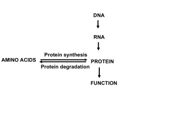
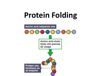
Proteins
L K Protein Structure W G …LWGK… Primary Alpha-helix Secondary Beta chain of Hemoglobin Tertiary Hemoglobin Quaternary
Some Rules to ‘Fold’ a Protein l Covalent interactions l Protein Sequence l Di-sulfide bridges l Non-covalent interactions l Hydrophobic interactions l Hydrogen bonds l Salt bridges (positive negative interactions) l Metal coordination
Protein Modeling: Toober and Thumb Tacks model l 1Toober l 10 thumb tacks l 1 Blue (Basic) l 1 Red (Acidic) l 4 Yellow (Hydrophobic) l 2 White (Hydrophilic) l 2 Green (Cysteine) http://www.3dmoleculardesigns.com/15_Tacks.pdf
Interaction rules l Hydrophobic (yellows should be away from water, and whites should be near water) l Charge based (red and blue should pair up) l Disulphide (the greens should pair up to form a bond)
PDB & Protein Models Protein Structures Protein Data Bank Protein Modeling
shown: the ribosome Why Structure? l Allows you to “visualize” the shape and details of the protein l Offers clues about the role in the body l May hold key to developing new medicines and diagnostic procedures for diseases like avian flu, HIV, West Nile Virus, parts of the protein associated with Alzheimer’s disease, Cancers, etc.
Understanding Protein Structures X-ray (X-ray crystallography) NMR EM (Nuclear Magnetic Resonance) (Electron Microscopy) Protein Data Bank Free download for use
The Protein Data Bank
The Structure Explorer Page
The PDB file
The PDB file – con’t.
Visualization (RasMol or Jmol)
Structure Representation Wireframe Ribbons Spacefill
Graphics KiNG WebMol Jmol Protein Explorer Default image
Zinc Finger Toober Model l Download the 1ZAA pdb file (www.pdb.org) l Create image in Jmol, identify key features l Fold a Mini-Toober model l Material modifications l Blue thumb tack (N-terminus) l Red thumb tack (C-terminus) l Colored Pipe-cleaners to represent Cys, His, Arg18, Phe16 and Leu22
NJSO 2008 Basic information
Information l Regional and State level contests l Protein Calmodulin (PDB ID 1CLL) l Rules l Pre-build: Bring in toober model and short abstract l On-site build: Build a designated part of 1CLL using a mini-Toober and Jmol l On-site exam: Answer questions about structure, function, importance and history of modeled protein. Materials will be provided.
Molecule of the Month
Structure Explorer Page
Jmol
How we judge Rubrics available from education.pdb.org/olympiad
Help l Details and links at http://education.rcsb.org/olympiad/ l If you have questions or to borrow the “I ntroduction to Protein Structure” collection suitcase please write to buildmodel@rcsb.rutgers.edu
Recommend
More recommend
Unleash a World of Digital Possibilities—Browse, Share, and Explore Content Without Boundaries