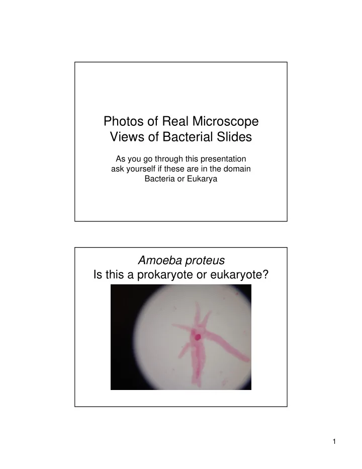

Photos of Real Microscope Views of Bacterial Slides As you go through this presentation ask yourself if these are in the domain Bacteria or Eukarya Amoeba proteus Is this a prokaryote or eukaryote? 1
Amoeba proteus at a higher magnification Nostoc slide 2
Nostoc at higher magnification Bacillus anthracis an example of a steptobacillus 3
Bacillus subtilis with endospores The yellow part inside the bacillus is an endospore Bacillus cell shape 4
Bacillus cell shape Bacillus with peritrchous flagella 5
Bacillus with peritrchous flagella at a higher magnification Large clumps of poorly focused cocci Focus should have been concentrated in on the orange circle. 6
Micrococcus luteus better focus of cocci; remember cocci arrange into packets of four cells, called tetrads. Poorly focused typical spirillum bacterial cell shape 7
Rhodospirillum rubrum a spirillum cell shape Euglena gracialis 8
Paramecium caudatum Paramecium caudatum 9
Saccharomyces cerevisiae Saccharomyces cerevisiae 10
Recommend
More recommend