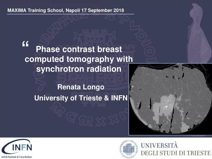

MAXIMA Training School, Napoli 17 September 2018 “ Phase contrast breast computed tomography with synchrotron radiation ” Renata Longo University of Trieste & INFN
MAXIMA Training School, Napoli 17 September 2018 “ Phase contrast breast computed tomography with synchrotron radiation ” Renata Longo University of Trieste & INFN
Characteristics of synchrotron radiation High X-ray intensity on a broad energy range – Tunable monochromatic beam Laminar beam geometry: the beam is naturally collimated – Images are acquired by scanning the object through the fan beam Small source size and large source-to-sample distance – High degree of lateral coherence Optimal X-rays source for phase contrast imaging Trieste, 24 September, 2018 - 3
Characteristics of synchrotron radiation Di EPSIM 3D/JF Santarelli, Synchrotron Soleil - Synchrotron Soleil, Attribution, https://commons.wikimedia.org/w/index.php?curid=376907 Trieste, 24 September, 2018 - 4
The SYRMEP Beamline (I) at Elettra “ detector sample patient monochromator detector source filters bending slit systems magnet experimental patient room hutch Bending Magnet Source Source size ~ 1.1 (horizontal) x 0.1 (vertical) mm 2 Monochromatic beam with tuneable energy (8.5 - 38 keV) Bandwidth: Dl/l ~ 2 x 10 -3 ” Divergence: ~ 7 mrad (horizontal) x 0.2 mrad (vertical)
The SYRMEP Beamline (II) “ detector sample monochromator patient detector source filters slit systems experimental patient room hutch Source-to-Sample distance: ~ 23 m ~32 m Laminar beam cross section: 4 x 150 mm 2 4 x 210 mm 2 Flux available at 17 keV (Elettra operated at 2.4 GeV, 140 mA ring current): 6 10 8 ph/mm 2 /s 2 10 8 ph/mm 2 /s ” Transverse coherence length 8 µm 11 µm
MAXIMA Training School, Napoli 17 september 2018 “ Phase contrast breast computed tomography with synchrotron radiation Credits Ioannis Sechopoulos Radboudumc ” Renata Longo University of Trieste & INFN
Breast CT “ 1 cm http://www.auntminnie.com credits J. Boone ”
Breast CT “ http://koninghealth.com/en/kbct/ 1 cm ” Bushberg et al, The essential physics of medical imaging
Breast CT @ SYRMEP “ ”
Breast CT @ SYRMEP Single Photon Counting Detector Large area CdTe single photon counting detector (PIXIRAD-8) PROS - Need for multi- - High efficiency module architecture - Large area (25x2.5 cm2) (3 pixel gap between - Small pixel (~60µm) modules) direct conversion - Time/exposure dependent charge - Dead Time Free Mode trapping effects - Tunable Threshold A dedicated pre- - No electronic noise CONS processing has been implemented! Bellazzini, R., et al. JINST 8.02 (2013): C02028. Delogu, P., et al. JINST 11.01 (2016): P01015. Delogu, P., et al. JINST 12.11 (2017): C11014. Brombal L. et al JSR (2018) Courtesy of Luca Brombal Trieste, 24 September, 2018 - 11
Preprocessing effectiveness “ WITHOUT PREPROCESSING Breast specimen: Beam energy 32 keV, MGD 20mGy WITH PREPROCESSING 1 cm 1 cm ” Brombal L et al JSR 2018 Luca Brombal
Preprocessing effectiveness “ WITHOUT PREPROCESSING Breast specimen: Beam energy 32 keV, MGD 20mGy WITH PREPROCESSING Brombal L et al JSR 2018 ” 1 cm 1 cm FisMat 2017, Trieste, 1-5 October 2017 Luca Brombal 12
Optimization of the threshold “ CdTe sensor E =26 KeV Pixirad I/ Pixie-III Di Trapani et al JINST-proceedings IWORID 2018 ”
Optimization of the threshold “ CdTe sensor E = 33 KeV Pixirad I/ Pixie-III Di Trapani V. et al JINST-proceedings IWORID 2018 ”
Linearity and threshold “ Delogu P. et al 2016 JINST 11 P01015 ”
MAXIMA Training School, Napoli 17 september 2018 “ Phase contrast breast computed tomography with synchrotron radiation ” Renata Longo University of Trieste & INFN
Phase-Sensitive Imaging Techniques “ The principle is the phase shift f (x,y) of the X-ray wave f (x,y) ~ 10-100 m rad • A family of techniques has been developed to transform phase shift into intensity modulation on the detector Zhou, S.-A. and Brahme, A. (2008). • Refraction index for hard X-rays n = 1 - d + i b Phys. Med. 24 , 129 – 148 phase shift absorption ”
Absorption imaging and “ Propagation-based Phase-Contrast Imaging Incoming Transmitted Absorption Position object Intensity Incoming Transmitted Position a object ” Intensity 10< a <100 m rad
A clinical study in phase contrast mammography “ 71 patients CONV PCI ”
Synchrotron radiation for medical imaging • Refraction index for hard X-rays 1000 𝒐 = 𝟐 − 𝜺 + 𝒋𝜸 100 phase shift absorption 10 d / b • For low Z elements ( 𝜺 >> 𝜸 ) 1 phase effects >> absorption 0.1 100 1000 10000 energy [eV] • In free-propagation the phase contrast is proportional to the second derivative of the phase shift ( ∇ 2 Φ (x,y )) → edge enhancement Need for a (partially) coherent source (e.g., the synchrotron) Trieste, 24 September, 2018 - 21
An exercise: the shell “ What are acquired at high energy? What are acquired in phase contrast ? ”
From phase contrast to phase retrieval • The phase signal can be extracted from a single Phase contrast phase contrast image using a phase retrieval algorithm (and suitable approximations) A phase-retrieved image is obtained • The used phase retrieval algorithm is based on the homogeneous Transport of Intensity Equation (TIE-Hom) and 𝜺, 𝜸 are assumed to be proportional • From a signal processing perspective, phase Phase retrieved retrieval is a low-pass filter in 2D Fourier domain applied to the projections λ = is the wavelength, w = (u,v) = spatial frequency Paganin, D. et al. Journal of microscopy 206.1 d = the propagation distance (2002): 33-40. Trieste, 24 September, 2018 - 23
… More on the phase retrieval … Gureyev, T.E., et al. JOSA A 34.12 (2017): 2251-2260. • CNR does not change upon free space propagation (in the near-field region) while spatial resolution improves (i.e., high spatial frequencies are enhanced) • CNR increases upon TIE-Hom (phase) retrieval . Spatial resolution deteriorates upon TIE-Hom retrieval (low pass filter), back to the level it had in the contact plane. The net effect is that the ratio of CNR and spatial HERE THE MAGIC HAPPENS! resolution increases in TIE-HOM imaging Trieste, 24 September, 2018 - 24
An exercise: a breast CT Before or after phase-retrieval ???? 1 cm @32 KeV 20 mGy MGD Trieste, 24 September, 2018 - 25
Single and double materials approaches • Assuming an homogeneous object (i.e., breast equivalent tissue) the PhR filter is: Single material • Considering interfaces between 2 materials (i.e., fat/glandular tissue interface) the PhR is: Double material Burvall, Anna, et al. Optics express 19.11 (2011) Spatial From a signal processing perspective higher CNR Resolution 𝜺/𝜸 narrower the filter in the Fourier domain (𝜺 𝟐 − 𝜺 𝟑 )/(𝜸 𝟐 − 𝜸 2 ) 𝜺/𝜸 869 2302 @32 keV Trieste, 24 September, 2018 - 26
PHASE RETRIEVED (DOUBLE MATERIAL) NO PHASE RETRIEVAL FWHM~190µm FWHM~120 µ m CNR ~ 3.5 CNR ~ 0.7 1 cm @32 KeV 20 mGy MGD Brombal L et al SPIE Medical Imaging 2018 Trieste, 24 September, 2018 - 27
Is phase-retrieval useful ? NO PHASE RETRIEVAL PHASE RETRIEVED 2 mm 170 mGy 20 mGy 5 mGy @32 keV MGD Trieste, 24 September, 2018 - 28
Optimization 3 m 9 m 1.6 m Higher the propagation distance better the image propagation distance Gureyev, T.E., et al. JOSA A 3(2017), Brombal L. et al PMB (submitted) Trieste, 24 September, 2018 - 29
5 minutes break ? “ • Questions • Comments • A little walk … ”
SYRMA-3D collaboration “ To develop methods and the facility to perform in phase-contrast (PhC) breast computed tomography (breast-CT) exploiting the monochromatic beam available at Elettra synchrotron radiation (SR) laboratory in Trieste in Italy Completed the first SR mammography clinical study: in our 71 patients PhC SR mammography outperforms conventional digital mammography (DM) ” Fedon, C. et al. J. Med. Imag. 5(1), 013503 (2018)
Clinical exam requirements • SAFETY & • IMAGING DOSIMETRY High resolution Low Delivered dose High CNR Strict exam Large Scanned protocol volume Fast Comfortable reconstruction patient table Well established Reasonable data workflow exam duration Safe data storage • COMFORT • DATA Trieste, 24 September, 2018 - 32
Methods and Materials “ Images large breast specimens were acquired at the SR facility used for the clinical study of PhC mammography and up-dated for breast-CT Longo R. et al Phys Med Biol (2016) The monochromatic x-ray beam was set in the energy range 30-38 keV and the delivered mean glandular dose (MGD) was in the range 5-20 mGy Mettivier G. et al Phys Med Biol (2016) The propagation-based PhC imaging technique was used and the phase-retrieval algorithm was applied before the FBP reconstruction Paganin D. et al. Journal of microscopy (2002) ”
Low-dose 3D images E = 32 keV MGD= 5 mGy Courtesy of Sandro Donato Trieste, 24 September, 2018 - 34
Recommend
More recommend