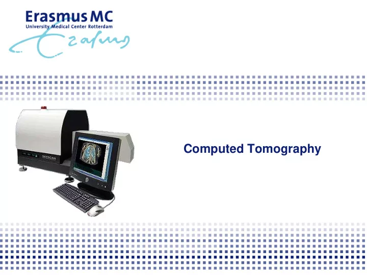

Computed Tomography
Outline X-RAY Computed Tomography Artifacts and Sources of Error Imaging studies Bone Cartilage Other
Outline X-RAY Computed Tomography Artifacts and Sources of Error Imaging studies Bone Cartilage Other
X-RAY: radiation type X-rays are ionizing, invisible and highly penetrating electromagnetic radiation
X-RAY: how to generate? Heated filament Electrons are emits electrons by accelerated by thermionic emission a high voltage X-RAY produced when high speed electrons hit metal target
X-RAY: attenuation Attenuation depends on: Thickness Density Effective atomic number Energy of the X-RAY beam
X-RAY Projection: Projections over 180 o Reconstruct to 3D Fast Non-destructive Direct 3D calculations
Outline X-RAY Computed Tomography Artifacts and Sources of Error Imaging studies Bone Cartilage Other
CT – Back Projection In standard back-projection, images are taken for a number of angles positions and then smeared back to form the section
CT – Back Projection Original Back projection Shadow
Outline X-RAY Computed Tomography Artifacts and Sources of Error Imaging studies Bone Cartilage Other
Artifacts and Sources of Error Radiation dose 7 Total exposure in mGy in mice: 6 5 4 3 2 1 0 0.0 10.0 20.0 30.0 40.0 50.0 Scan resolution (microns)
Artifacts and Sources of Error Beam hardening: Theoretical model: beam with only high energy photons 20% 35% 50% 60% 20% 35% High energy photons Low energy photons
Artifacts and Sources of Error Beam hardening: Beam hardening: Theoretical model: beam with only high energy photons 35% 20% 60% 50% 35% 20% True model: 35% 75% error mixed beam 60% 20% error 35% High energy photons Low energy photons
Artifacts and Sources of Error Rat femur with titanium implant:
Artifacts and Sources of Error Beam hardening, correction via: Physical filter (aluminum) Correction afterwards using software Unfiltered Filtered
Artifacts and Sources of Error Ring artifacts: due to drift and nonlinearities in the response of individual elements in the detector
Artifacts and Sources of Error Alignment error:
Outline X-RAY Computed Tomography Artifacts and Sources of Error Imaging studies Bone Cartilage Other
Micro-CT analysis 3D-models: Lateral Medial Trabecular bone Epiphysis Cross-section
Bone Control Osteoarthritis induced
Bone Architectural bone changes validated with histology New bone Resorbed bone
Bone Femur defect Filled with porous titanium structure
Outline X-RAY Computed Tomography Artifacts and Sources of Error Imaging studies Bone Cartilage Other
uCTa: microCT-arthrography Synovial fluid Healthy microCT Cartilage Bone
uCTa: microCT-arthrography Synovial fluid Healthy microCT Cartilage Bone
uCTa: microCT-arthrography Synovial fluid OA microCT Cartilage Bone
Contrast-enhanced microCT Healthy OA-induced / mono-iodoacetate control Low attenuation High attenuation (high GAG) (low GAG)
Micro-CT analysis
Outline X-RAY Computed Tomography Artifacts and Sources of Error Imaging studies Bone Cartilage Other
Contrast-enhanced microCT Mouse lung sample With contrast agent 5.7 μ m pixel size Source: http://www.skyscan.be/applications/biomedical/biomed002.htm
Contrast-enhanced microCT Mouse kidney sample With contrast agent 80 μ m pixel size Source: http://www.scanco.ch/applications/biomedical.html
Contrast-enhanced microCT Rat vascular system With contrast agent 75 μ m pixel size vivaCT Source: http://www.scanco.ch/applications/biomedical.html
Cardiac function Cranial Caudal Diastole Systole
Contrast-enhanced microCT Mouse (in-vivo) NO contrast agent 160 μ m pixel size Source: http://www.skyscan.be/applications/biomedical/biomed006.htm
Contrast-enhanced microCT Mouse (in-vivo) WITH contrast agent 160 μ m pixel size Real-time radiography of heart activity and contrast propagation Source: http://www.skyscan.be/applications/biomedical/biomed006.htm
Recommend
More recommend