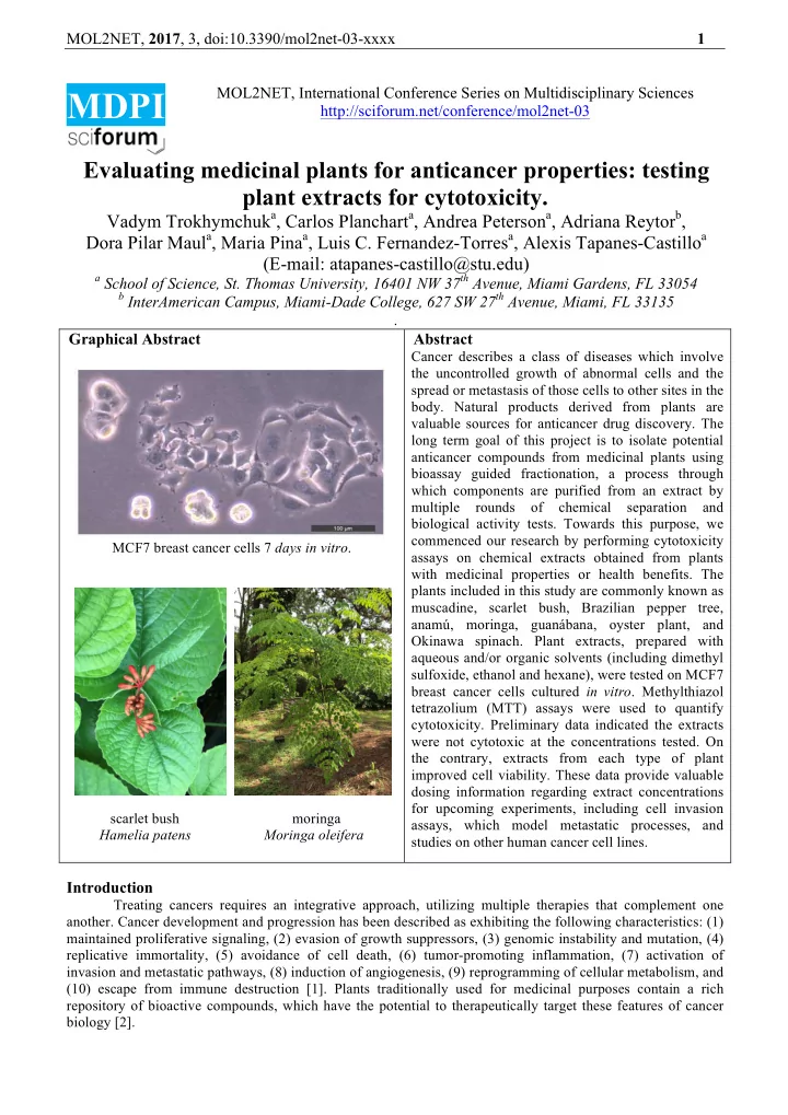

MOL2NET, 2017 , 3, doi:10.3390/mol2net-03-xxxx 1 MOL2NET, International Conference Series on Multidisciplinary Sciences MDPI http://sciforum.net/conference/mol2net-03 Evaluating medicinal plants for anticancer properties: testing plant extracts for cytotoxicity. Vadym Trokhymchuk a , Carlos Planchart a , Andrea Peterson a , Adriana Reytor b , Dora Pilar Maul a , Maria Pina a , Luis C. Fernandez-Torres a , Alexis Tapanes-Castillo a (E-mail: atapanes-castillo@stu.edu) a School of Science, St. Thomas University, 16401 NW 37 th Avenue, Miami Gardens, FL 33054 b InterAmerican Campus, Miami-Dade College, 627 SW 27 th Avenue, Miami, FL 33135 . Graphical Abstract Abstract Cancer describes a class of diseases which involve the uncontrolled growth of abnormal cells and the spread or metastasis of those cells to other sites in the body. Natural products derived from plants are valuable sources for anticancer drug discovery. The long term goal of this project is to isolate potential anticancer compounds from medicinal plants using bioassay guided fractionation, a process through which components are purified from an extract by multiple rounds of chemical separation and biological activity tests. Towards this purpose, we commenced our research by performing cytotoxicity MCF7 breast cancer cells 7 days in vitro . assays on chemical extracts obtained from plants with medicinal properties or health benefits. The plants included in this study are commonly known as muscadine, scarlet bush, Brazilian pepper tree, anamú, moringa, guanábana, oyster plant, and Okinawa spinach. Plant extracts, prepared with aqueous and/or organic solvents (including dimethyl sulfoxide, ethanol and hexane), were tested on MCF7 breast cancer cells cultured in vitro . Methylthiazol tetrazolium (MTT) assays were used to quantify cytotoxicity. Preliminary data indicated the extracts were not cytotoxic at the concentrations tested. On the contrary, extracts from each type of plant improved cell viability. These data provide valuable dosing information regarding extract concentrations for upcoming experiments, including cell invasion scarlet bush moringa assays, which model metastatic processes, and Hamelia patens Moringa oleifera studies on other human cancer cell lines. Introduction Treating cancers requires an integrative approach, utilizing multiple therapies that complement one another. Cancer development and progression has been described as exhibiting the following characteristics: (1) maintained proliferative signaling, (2) evasion of growth suppressors, (3) genomic instability and mutation, (4) replicative immortality, (5) avoidance of cell death, (6) tumor-promoting inflammation, (7) activation of invasion and metastatic pathways, (8) induction of angiogenesis, (9) reprogramming of cellular metabolism, and (10) escape from immune destruction [1]. Plants traditionally used for medicinal purposes contain a rich repository of bioactive compounds, which have the potential to therapeutically target these features of cancer biology [2].
MOL2NET, 2017 , 3, doi:10.3390/mol2net-03-xxxx 2 Materials and Methods Table 1: Plant extract concentrations tested MCF7 breast cancer cells (American Type Extract mg/mL Culture Collection) were plated on 96-well plates at a Control (DMSO) 110 or 154 density of 15,000-20,000 cells per well. Cells were Anamú Leaves 7.1 cultured in Dulbecco’s Modified Eagle Anamú Roots 9.4 Medium/Nutrient Mixture F-12, 10% fetal bovine Braz. Pepper Bark 75 EtOH/25 Hex 0.1 serum, and 1X penicillin/streptomycin. Braz. Pepper Bark (dried) 1.9 Plant extracts were prepared from muscadine Braz. Pepper Berry (dried) 9.4 ( Vitis rotundifolia ), scarlet bush ( Hamelia patens ), Braz. Pepper Berry 100 EtOH 8.8 Brazilian pepper tree ( Schinus terebinthifolius ), anamú Braz. Pepper Berry 75 EtOH/25 Hex 2.4 ( Petiveria alliacea ), moringa ( Moringa oleifera ), Braz. Pepper Leaves (dried) 9.4 guanábana ( Annona muricata ), oyster plant Braz. Pepper Leaves 75 EtOH/25 Hex 3.3 ( Tradescantia spathacea ), and Okinawa spinach Braz. Pepper Leaves 50 EtOH/50 Hex 20.0 ( Gynura bicolor ) utilizing aqueous and/or organic Braz. Pepper Leaves CH2Cl2 0.9 solvents [3-10]. Dimethyl sulfoxide (DMSO) was Guanábana Leaves 1.0 added to extracts at the concentrations listed for the Moringa Bark 1.5 control (Table 1) to improve solubility and cellular Moringa Leaves 1.5 internalization. Extracts were then filter-sterilized and Moringa Seeds 9.3 diluted in media as described in Table 1. Muscadine Fruit (dried) 1.4 Concentrations varied between extracts because the aim Muscadine Fruit (whole) 1.4 was to maximize extract concentration, not to test Muscadine Fruit Outer Shell 39.5 uniform extract concentrations. Muscadine Fruit Pulp 28.3 Cells were first cultured for 48 hours and then Muscadine Leaves 100 EtOH 13.3 treated with plant extracts for 72 hours. Methylthiazol Muscadine Leaves 75 EtOH/25 Hex 10.0 tetrazolium (MTT) assays were conducted to evaluate cytotoxicity. Culture media was replaced with Muscadine Leaves 50 EtOH/50 Hex 7.8 RPMI1640 (without phenol red), 10% fetal bovine Muscadine Leaves CH2Cl2 3.6 serum, and 0.5 mg/mL MTT. Formazan crystals were Muscadine Roots 75 EtOH/25 Hex 11.6 solubilized in 0.1 N HCl (diluted in isopropanol). Oki. Spinach Leaves Aqueous 9.3 Samples were read on a Synergy H1 (BioTek) plate Oyster Plant Leaves 50 EtOH/50 Hex 3.6 reader set to 570 nm with a 630 nm reference Scarlet Bush Leaves 61.7 background subtraction. Each extract was tested using 5-16 replicates in one or two independent experiments. Data from extract- treated cells were compared to that obtained from untreated controls with the same DMSO concentration. Absorbance values were averaged and normalized to controls. Standard deviations of normalized values were calculated. Two-sample, two-tail t -tests assuming unequal variances were utilized to calculate p -values. In the case of multiple experiments, the largest p -value was reported. Results and Discussion Figure 1 summarizes preliminary data regarding the effect of plant extracts on MCF7 breast cancer cells. Extracts were not cytotoxic at the concentrations tested. The greater the absorbance, the higher the concentration of formazan, a purple product generated in live cells by mitochondrial succinate dehydrogenase (SDH) enzyme reduction of MTT. Variability in the data, especially evident in the larger error bars observed at higher values, are primarily attributed to the challenge of consistently solubilizing higher formazan concentrations with manual trituration. Wells treated with extracts from each type of plant had significantly higher SDH activity during the MTT assay than untreated wells (Fig. 1). This activity is proportional to the number of live cells in a well, which is related to cell survival and cellular proliferation. Hence, overall the extracts reduced cell death. These results are not surprising given that extracts from these plants have been reported to have medicinal effects [reviewed in 3- 8]. Moreover, recent studies suggest several of the extracts demonstrate antioxidant activity in chemical reactions [3-8]. In general, antioxidants improve cell viability, including that of cancer cells, which tend to exhibit elevated levels of reactive oxygen species due to metabolic and signal transduction aberrations related to tumorigenesis [11]. Antioxidants provided by the plant extracts could reduce electron leakage during mitochondrial respiration and superoxide formation, increasing the number of live cells in treated samples.
Recommend
More recommend