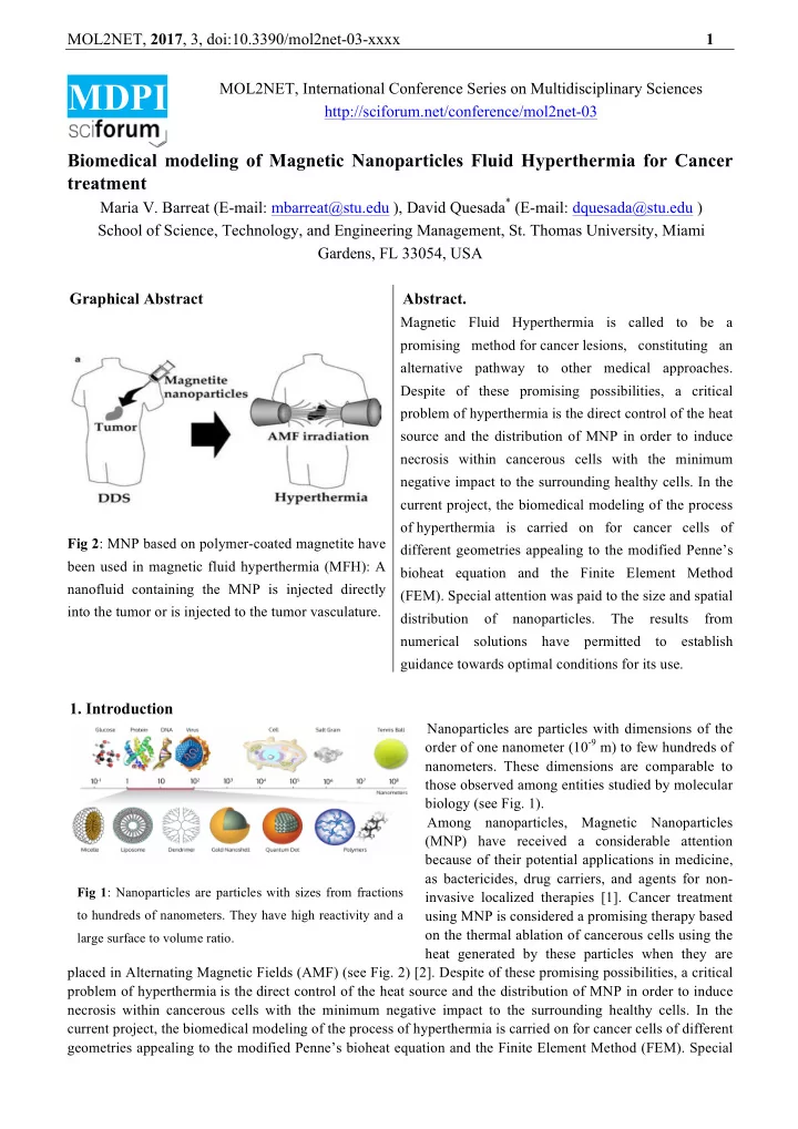

MOL2NET, 2017 , 3, doi:10.3390/mol2net-03-xxxx 1 MDPI MOL2NET, International Conference Series on Multidisciplinary Sciences http://sciforum.net/conference/mol2net-03 Biomedical modeling of Magnetic Nanoparticles Fluid Hyperthermia for Cancer treatment Maria V. Barreat (E-mail: mbarreat@stu.edu ), David Quesada * (E-mail: dquesada@stu.edu ) School of Science, Technology, and Engineering Management, St. Thomas University, Miami Gardens, FL 33054, USA Graphical Abstract Abstract. Magnetic Fluid Hyperthermia is called to be a promising method for cancer lesions, constituting an alternative pathway to other medical approaches. Despite of these promising possibilities, a critical problem of hyperthermia is the direct control of the heat source and the distribution of MNP in order to induce necrosis within cancerous cells with the minimum negative impact to the surrounding healthy cells. In the current project, the biomedical modeling of the process of hyperthermia is carried on for cancer cells of Fig 2 : MNP based on polymer-coated magnetite have different geometries appealing to the modified Penne’s been used in magnetic fluid hyperthermia (MFH): A bioheat equation and the Finite Element Method nanofluid containing the MNP is injected directly (FEM). Special attention was paid to the size and spatial into the tumor or is injected to the tumor vasculature. distribution of nanoparticles. The results from numerical solutions have permitted to establish guidance towards optimal conditions for its use. 1. Introduction Nanoparticles are particles with dimensions of the order of one nanometer (10 -9 m) to few hundreds of nanometers. These dimensions are comparable to those observed among entities studied by molecular biology (see Fig. 1). Among nanoparticles, Magnetic Nanoparticles (MNP) have received a considerable attention because of their potential applications in medicine, as bactericides, drug carriers, and agents for non- Fig 1 : Nanoparticles are particles with sizes from fractions invasive localized therapies [1]. Cancer treatment to hundreds of nanometers. They have high reactivity and a using MNP is considered a promising therapy based on the thermal ablation of cancerous cells using the large surface to volume ratio. heat generated by these particles when they are placed in Alternating Magnetic Fields (AMF) (see Fig. 2) [2]. Despite of these promising possibilities, a critical problem of hyperthermia is the direct control of the heat source and the distribution of MNP in order to induce necrosis within cancerous cells with the minimum negative impact to the surrounding healthy cells. In the current project, the biomedical modeling of the process of hyperthermia is carried on for cancer cells of different geometries appealing to the modified Penne’s bioheat equation and the Finite Element Method (FEM). Special
MOL2NET, 2017 , 3, doi:10.3390/mol2net-03-xxxx 2 attention was paid to the size and spatial distribution of nanoparticles. The results from numerical solutions have permitted to establish guidance towards optimal conditions for its use. 2. Mathematical model and Numerical methods In order to simulate the heating of cancer cells the modified Penne’s bioheat equation [3] is used. It is a heat- type equation complemented with terms responsible for the heat exchange between the cell and blood, and also the metabolic heat and the one produced by the embedded MNP. Equations can be cast into: ∂ T ! k ! ∇ ! T ! + ρ ! c ! ω ! T ! − T ! + Q ! + P ρ ! c ! = ∂ t ∂ T ! k ! ∇ ! T ! + ρ ! c ! ω ! T ! − T ! + Q ! ρ ! c ! = ∂ t where the sub indices t and h refer to tumor and healthy cells respectively, ρ is the tissue density, c is the specific heat for the tissue, k is the thermal conductivity, ρ b is the blood density, c b is the specific heat of the blood, ω t ( ω h ) is the blood perfusion rate, and T b is blood temperature. The terms Q t and Q h are metabolic sources of heat for tumor and healthy cells respectively. The function P(T,H) is the rate of heat production by the MNP and includes the information about the static and dynamic components of the power dissipated by MNP. It is given by the following equation: P T , H = 1 ω 𝜐 !"" ! ω 2 µ ! χ ! H ! 1 + ( ω 𝜐 !"" ) ! where H 0 is the intensity of the applied magnetic field, ω is the frequency of the AC applied magnetic field, µ 0 is the magnetic permeability of the vacuum, χ 0 is the static component of the magnetic susceptibility, τ eff is the effective relaxation time of MNP due to Brown (rotation of MNP in the viscous medium) and Neel (rotation of magnetic moments) mechanisms. The explicit mathematical expressions are: 𝐿𝑊 3 𝜃 𝑊 ! ! + 𝜐 ! ! ! ! ; 𝜐 !"" = 𝜐 ! 𝜐 ! = 𝜐 ! ex p 𝑙 ! 𝑈 ; 𝜐 ! = 𝑙 ! 𝑈 ! 𝑊 3 𝜊 coth 𝜊 − 1 𝜈 ! 𝜚 𝑁 ! 𝜈 ! 𝑁 ! 𝐼𝑊 𝜓 ! ( 𝑈 , 𝐼 ) = 𝜓 ! 𝜊 ; 𝜓 ! = ; 𝜊 = 3 𝑙 ! 𝑈 𝑙 ! 𝑈 where η is the dynamic viscosity of a medium where particles are suspended, K is the effective anisotropy constant, V H is the particle hydrodynamic volume, V is the volume of the magnetic core, M d is the domain magnetization of the MNP, and ϕ is the volume fraction solid. 3. Results and Discussion The parameters used for simulations are summarized in tables 1 and 2. Tumor Healthy Tissue Blood Density – ρ (kg/m 3 ) 1045 1045 1060 Heat capacity – c (J/kg K) 3760 3760 3770 Thermal conductivity – k (W/m K) 0.51 0.51 ---- Blood perfusion rate – ω (1/s) 0.0095 0.003 ---- Metabolic heat – Q (W/m 3 ) 31872.5 6374.5 ---- Table 1 : Physical parameters characterizing each medium that were used in simulations. Magnetic NP radius R p = 9.5 x 10 -9 m k B = 1.38 x 10 23 J/K Volume fraction solid ϕ = 0.071 Effective anisotropy constant K = 1.0 x 10 4 J/m 3 Dynamic viscosity η = 1.0 x 10 -3 kg/m s f = 300 kHz Hydrodynamic volume V H = 5.08 x 10 -22 m 3 Attempt time τ 0 = 10 -9 s µ 0 = 4 π x 10 -7 T m/A Domain magnetization M d = 446 kA/m Field strength H 0 = 5518 A/m Table 2 : Physical parameters and constants used in simulations. The above system of partial differential equations (PDE) was discretized according to the Finite Differences and solved numerically following the Crank-Nicholson method. The inclusion of the function P(T,H) transforms the system from linear to non-linear, which demands a more careful attention. Solutions were found and plotted
MOL2NET, 2017 , 3, doi:10.3390/mol2net-03-xxxx 3 with Wolfram Mathematica (Figs 3 and 4). In Fig. 3 the static magnetic susceptibility has been computed including a convolution by the distribution of particle’s radii assumed to be Log-normal. Curves in red and blue correspond to particle’s radii of 9.46 and 9.11 nm respectively, while the green one corresponds to 10.30 but different standard deviation, making it to appear narrower than the other two. The choice for a Log-normal distribution is based on results from [2]. 0.25 10 -3 3.5 0.018 0.016 3 0.2 0.014 2.5 Cluster Magnetic Susceptibility 0.012 Particle Distribution 0.15 2 Susceptibility 0.01 0.008 1.5 0.1 0.006 1 0.004 0.05 0.5 0.002 0 0 0 0 5 10 15 20 25 30 35 40 0 5 10 15 20 25 30 35 40 0 5 10 15 20 25 30 35 40 Particle size in nm Particle size in nm Particle size in nm Fig 3 : Computation of the Probability Distribution Function (PDF) (left) for nanoparticles, the magnetics susceptibility (center), and the cluster susceptibility (right). The computation of the heat distribution was done for tumor cells of circular shape embeeded into a ractangular frame, as can be seen from Fig. 4. The plots show the ratio of the actual temperature to a characteristic temperature, in order the make the system of equations dimensionless. As the time goes the tumor cell is heating from the center, where the nanoparticle cluster was located. More irregular geometries are in progress and are more suitable for invasive tumor cells of rapid growth. Likewise, different nanoparticle cluster’s geometries are impacting the effectivenes of using MFH. 0.22123270000 0.93616047500 Fig 4 : Numerical solution of the 0.93616045000 0.22123265000 0.93616042500 Modified Pennes Bio-heat equation 0.22123260000 0.93616040000 for spherical cell model. The 0.22123255000 0.93616037500 temperature profiles at two different moments are shown, noticing the gradual increase in temperature. 4. Conclusions The distribution of heat inside a tumor tissue has been computed and the effect of the distribution of MNP on the intensity of the heat was determined. The heat increases initially and then drops towards the boundary of the tumor tissue preventing neighbor healthy tissues of being affected. Acknowledgments Authors appreciate the support received from both, St. Thomas University and Miami Dade College, as wells as from the Department of Education grant P03C1160161 (STEM-SPACE). References 1. Luo S., Wang LF, Ding WJ, Wang H, Zhou JM, Jin HK, Su SF, et al. Clinical trials of magnetic induction hyperthermia for treatment of tumors, OA Cancer 18 , 1 – 6 (2014). 2. Salunkhe AB, Khot VM, Pawar SH. Magnetic hyperthermia with magnetic nanoparticles: A status review, Current Topics in Medicinal Chemistry 14, x – x (2014). 3. Wu L, Cheng J, Liu W, Chen X. Numerical analysis of electromagnetically induced heating and bioheat transfer for magnetic fluid hyperthermia, IEEE Transactions on Magnetics 51 , No 2 (2015); Lakhssassi A, Kengne E, Semmaoui H. Modified Pennes’ equation modeling bioheat transfer in living tissues: analytical and numerical analysis, Natural Science 2, 1375 – 1385 (2010).
Recommend
More recommend