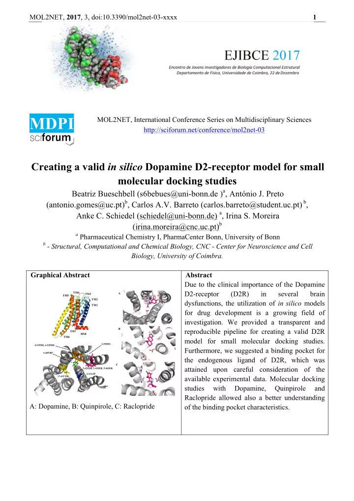

MOL2NET, 2017 , 3, doi:10.3390/mol2net-03-xxxx 1 MDPI MOL2NET, International Conference Series on Multidisciplinary Sciences http://sciforum.net/conference/mol2net-03 Creating a valid in silico Dopamine D2-receptor model for small molecular docking studies Beatriz Bueschbell (s6bebues@uni-bonn.de ) a , António J. Preto (antonio.gomes@uc.pt) b , Carlos A.V. Barreto (carlos.barreto@student.uc.pt) b , Anke C. Schiedel (schiedel@uni-bonn.de) a , Irina S. Moreira (irina.moreira@cnc.uc.pt) b a Pharmaceutical Chemistry I, PharmaCenter Bonn, University of Bonn b - Structural, Computational and Chemical Biology, CNC - Center for Neuroscience and Cell Biology, University of Coimbra. Graphical Abstract Abstract Due to the clinical importance of the Dopamine D2-receptor (D2R) in several brain TM4 A TM3 TM5 TM2 dysfunctions, the utilization of in silico models TM1 for drug development is a growing field of investigation. We provided a transparent and B reproducible pipeline for creating a valid D2R TM7 HX8 TM6 model for small molecular docking studies. 6.55HIS 6.51PHE, 6.52PHE Furthermore, we suggested a binding pocket for 6.48TRP the endogenous ligand of D2R, which was C attained upon careful consideration of the 5.42SER, 5.43SER, 5.46SER 3.32ASP available experimental data. Molecular docking (7.43TYR) studies with Dopamine, Quinpirole and Raclopride allowed also a better understanding A: Dopamine, B: Quinpirole, C: Raclopride of the binding pocket characteristics.
MOL2NET, 2017 , 3, doi:10.3390/mol2net-03-xxxx 2 Introduction Dopamine D2-receptor (D2R) is a member of G-Protein Coupled Receptors (GPCR) super-family, consisting in seven transmembranar helices (TM), three extracellular and three intracellular loops (1,2). D2R, along with D3R, are believed to have a more complex function within the dopamine receptor family as they are present in post- and pre-synaptic termina (3). In addition they exert a negative feedback loop in the presynaptic terminal to control firing rate of neurons (4). Along with this functional complexity, it has been shown that alterations in dopaminergic transmission in the brain are correlated to several dysfunctions such as Parkinson’s disease and Schizophrenia (5). These distinctive characteristics make D2R an important candidate for drug targeting and in silico drug development (3,5). Although some molecular docking studies have been performed with D2R, there are still a lot of problems and open questions that need a new effort in order to be fully understood. First, there is much incongruence in the way new models are created as the use of different templates and modeling softwares makes it hard for a clear comparison between models attained in different research groups. Second, absolute scores of model evaluation programs are often not shown, making model-building not transparent and comparable enough. Another problem rises from the various binding pockets definitions found in literature. Here, we tried to overcome this issue by using a large set of experimental data from Floresca et al (6). We aimed to attain a reproducible pipeline to calculate and evaluate correctly GPCR-models in general, and to D2R in particular. Materials and Methods Modelling The building of the D2R model was performed using MODELLER (7), with D3R complexed with Eticlopride, (Protein DataBank (8) ID 3PBL) as template (9). This crystallographic structure was chosen in accordance with a total sequence similarity of 68%, as calculated with BLAST (10). In addition each TM was then aligned to the TMs of the template, which numeration was obtained at the GPCRdatabase (11). The TMs were checked for sequence similarity and a average value of 77 % was attained for this more relevant and conserved helical bundle. Modelling was performed by specifying the lengths of the TMs and the perimembrane intracellular helix (HX8). Furthermore, disulphide bonds were considered in the pairs of unconserved cysteines at positions 79-154 and 249-251. Further loop refinement was performed when needed, in particular for the extracellular loop 2 (ECL2), since it is a long loop highly determinant for D2R’s binding pocket access. Since it is known that the deletion of the intracellular loop (ICL3) middle residues does not affect ligand and G-protein binding (the contact points are normally the N- and C-termini of this loop), residues 214-254 from ICL3 were removed as well as the first 28 residues of the sequence, as there is no template for these regions. A dialanine linker was added to connect TM5 and TM6, which were modelled as helices up to the linker, making the intracellular extension of TM5 and TM6 similar to what is observed in the G-protein bound crystal structure of the β 2 receptor (PDBid: 3SN6) (12). 2.50ASP was protonated as it known to interact with K+ binding site, which regulates by allostery the ions that regulate the function of the receptor. Furthermore, a part of the ICL2 (112-114) was set as alpha-helix in MODELLER protocol following the know structure-function experimental data available for this loop from the D3R (9). Model evaluation Although visual inspection immediately ruled out some trial models due to biological incoherence on particular and important structural features, selecting the best model was cumbersome. This was particularly true since D2R is a membrane protein, for which the more commonly available metrics are
MOL2NET, 2017 , 3, doi:10.3390/mol2net-03-xxxx 3 less reliable. Discrete Optimized Protein Energy (DOPE) (13) and molecules’ probability density functions (molpdf) are MODELLER’s standard metrics for model assessment, based on the models’ free energy and special occupation. These, however, did not allowed us to easily pick the best models, in part since they are mainly directed towards water soluble proteins. For this reason Protein Structure Analysis (ProSA) web service was additionally used for error recognition, in particular the z-score, which indicates overall model quality with respect to an energy distribution derived from random conformations (14). However, as the z-score was still not enough to choose the best model, we extended our analysis by using the online Protein Quality (ProQ) (15) prediction server. This is based on a neural network, the LGScore (16), to predict a p-value for the significance of a structural similarity match and the MaxSub (17) that identifies the largest subset of alpha carbons that superimposes with the template structure. Furthermore ProQ allows for the inclusion of secondary structure information (calculated by PSIPRED (18)) in order to further improve model quality assessment. Together with PSIPRED, ProQ can improve up to 15% its’ prediction accuracy. Finally, Ballesteros and Weinstein (19) numbering system for class A GPCRs was applied. The numbering system determines helical numbering (for TM1-7 and HX8) depending on a previously determined most conserved residue in each of the helices, named residue x.50. Docking Protocol AutoDockTools, a package of MGLTools was used to perform ligand docking (17). Docking itself was performed using Autodock4.2 (version autodock 4.2.6, released in 2009) (21). D2R hydrogens were added and Kollman united atom charges were assigned. Hydrogens were also added to ligand and Gasteiger-Marsili was used to calculate charges (22). Before docking an energy grid was created using Autogrid (version autogrid 4.2.6, released 2009) with a box-size of 50 Å x 50 Å x 50 Å in dimension with a 0.375 Å-spacing. The grid center was set at 18.235, 17.556, 6.595. For each docking simulation 100 independent Lamarckian genetic algorithm (LGA) runs were performed with the number of energy evaluations set to 10.000.000, the population size set to 200 and the maximum number of generations set to 27.000 (23). Default settings were maintained for the rest of the parameters. Docked conformations within a RMSD of 2 Å were clustered. The most populated and lowest energy cluster was used for conformational binding analysis. According to Floresca et al. (6), the following residues are important for ligand binding, and were therefore treated as fully flexible during the docking process, along with all rotatable bonds of ligands: 3.32ASP (ASP86), 5.42SER (SER165), 5.43SER (SER166), 5.46SER (SER169), 6.48TRP (TRP236), 6.51PHE (PHE239), 6.52PHE (PHE240), 6.55HIS (HIS243) and 7.43TYR (TYR266). In this study, we docked Dopamine, the endogenous ligand, Quinpirole, a selective D2R/D3R-agonist and Raclopride, a selective D2R-antagonist. We calculated all distances between the center of mass of the ligand and the alpha-C-atom (C α ) of the flexible residues used in the docking for all the top conformations achieved with AutoDock4.2 in order to attain an initial evaluation of these models (21). Results and Discussion Due to the high degree of homology between the D2R and D3R (total similarity 68%), it was suitable to use the recently solved crystal structure of the D3R as template (9,24). The ClustalOmega alignment showed that the chosen TMs for the D2R model were conserved compared to D3R (similarity for TM1: 60%, for TM2: 92.86%, for TM3: 87.88%, for TM4: 68.18%, for TM5: 64.71%, for TM6: 70.59%, for TM7: 84.00% and for HX8: 92.31%). For the D2R model a DOPE-score of -39622.63 and z-score of -2.55 (MODELLER and ProSA-web) were attained. In addition, the model was evaluated with ProQ with and without a PSIPRED secondary prediction LGscore: 3.43/3.78 and MaxSub: 0.12
Recommend
More recommend