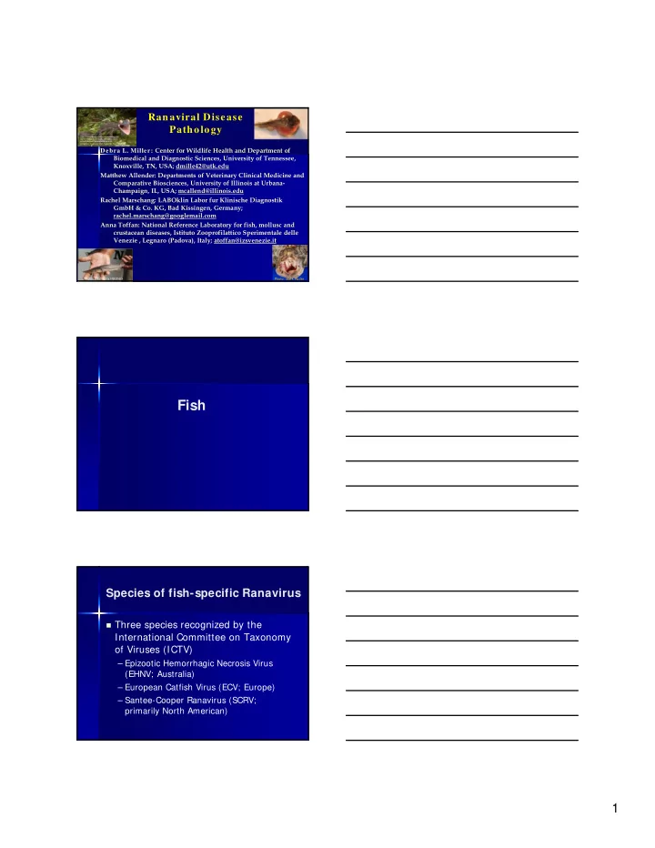

Ranaviral Disease Pathology Photo: N Haislip http://scienceblogs.com/tetrapodzoology/wp- content/blogs.dir/471/files/2012/05/i- ef0fe026ef8adf268fbce8dda99e3d45- Uroplatus_fimbriatus_Piotr-Naskrecki_April-2010.jpg Debra L. Miller: Center for Wildlife Health and Department of Biomedical and Diagnostic Sciences, University of Tennessee, Knoxville, TN, USA; dmille42@utk.edu Matthew Allender: Departments of Veterinary Clinical Medicine and Comparative Biosciences, University of Illinois at Urbana- Champaign, IL, USA; mcallend@illinois.edu Rachel Marschang: LABOklin Labor fur Klinische Diagnostik GmbH & Co. KG, Bad Kissingen, Germany; rachel.marschang@googlemail.com Anna Toffan: National Reference Laboratory for fish, mollusc and crustacean diseases, Istituto Zooprofilattico Sperimentale delle Venezie , Legnaro (Padova), Italy; atoffan@izsvenezie.it Photo: Blind Pony Hatchery Photo: Mark Ruder Fish Species of fish-specific Ranavirus Three species recognized by the International Committee on Taxonomy of Viruses (ICTV) – Epizootic Hemorrhagic Necrosis Virus (EHNV; Australia) – European Catfish Virus (ECV; Europe) – Santee-Cooper Ranavirus (SCRV; primarily North American) 1
….And some amphibian ranaviruses have been found to infect fish. FV3- – I n wild fish: moribund threespine stickleback ( Gasterosteus aculeatus ) during a sympatric epizootic involving northern red- legged frogs ( Rana aurora ; Mao et al. 1999a). – I n various hatcheries (e.g., Waltzek et al. 2014) BI V –only a single outbreak in hatchery-reared Nile tilapia fry ( Oreochromis niloticus ) in Australia (Ariel and Owens 1997 ). Ranaviruses in hatcheries FV3 and SCRV have been detected in various hatchery reared freshwater fishes in the Americas and Asia (see: Woodland et al. 2002b ; Prasankok et al. 2005 ; Deng et al. 2011 ; George et al. 2014 ; Chinchar and Waltzek 2014 ; Waltzek et al. 2014) Field and gross Loss of buoyancy Erratic swimming Anorexia Red swollen gills Hemorrhages (especially periocular, fat bodies, swim bladder) Overinflated swim bladder Friable (necrotic) organs 2
Fish Photo: Emilie Travis Photos: Tom Waltzek Photo: Ted Henry EHNV First ranavirus reported in mass die-off of vertebrate (Duffus et al. 2015) 1985, Australia, epizootic (see: Langdon et al. 1986, 1988; Langdon and Humphrey 1987); Unknown source of outbreak Redfin perch ( Perca fluviatilis ) and Rainbow trout ( Oncorhynchus mykiss ) Other species are susceptible based on laboratory challenges but no recent outbreaks (perhaps events aren’t detected) EHNV-current status Endemic in wild Redfin perch in SE Australia Impact to aquaculture = farmed rainbow trout in SE Australia Redfin perch = highly susceptible; Rainbow trout = relatively resistant (see: Whittington and Reddacliff 1995) 3
EHNV –field and gross Melanosis (dark color) Anorexic (stopped eating) Ataxic Swollen abdomen Swollen spleen and kidney Multifocal pale foci (areas of necrosis) in the liver See: Reddacliff and Whittington 1996 Gross lesions Multifocal hepatic necrosis and echymotic hemorrhage in the retroperitoneum; adult Redfin perch I mmunohistochemical staining in the areas of necrosis in the liver of a Redfin perch ( Perca fluviatilis ). Photo: Richard Whittington 4
ECV in European catfish by Anna Toffan (DVM, PhD) Aquatic Animal Virology Unit National Reference Laboratory for fish, mollusc and crustacean diseases Istituto Zooprofilattico Sperimentale delle Venezie (IZSVE) Viale dell’Università 10, 35020 Legnaro (Padova), Italy Tel 0039 049 8084333 Fax 0039 0498084360 email atoffan@izsvenezie.it Clinical signs • Mortality 100% • Melanosis (dark color) • Exophthalmos (pop eye) • Anorexia and lethargy • Eratic swimming, gasping , «candle» position • Petechial hemorrages on skin Clinical signs 5
Gross lesion • Hemorrhages on external and internal organs (skin, fins, bladder, intracoelomic fat, liver) • Anemic gill with petechiae and/or oedema • Congestion and protrusion of the anus • Congestion of the intestinal tract • Necrotic foci in liver, spleen, kindey • Spleen and liver enlargment Petechial hemorrhages Exophthalmos and eye hemorrhages 6
Intracoelomic (abdominal) hemorrhage Splenomegaly and petechial hemorrhages on liver Vascular congestion (especially on the stomach and intestines) 7
Histopathology Spleen: depletion of lymphoid tissue Liver: Necrotic foci with pyknosis of hepatocytes. pyknosis and cariorexis of white pulp Picture by Tobia Pretto ‐ IZSve IHC Kidney: Positive reaction in interstitial lymphoid tissue Picture by Tobia Pretto ‐ IZSve SCRV Typical die-off event – Fish die during summer – Often only largemouth bass ( Micropterus salmoides ) > 30 cm TL found – Moribund fish at water surface – External hemorrhages; however, there may be no external lesions unless there is another concurrent disease – Swim bladder is over-inflated, reddened, or has yellow or brown exudate – Fish-kill can last for 2-3 months 8
Photo: Ted Henry 9
SCRV Survives in water for several days Occurs in fish mucus (sometimes) Isolated from trunk kidney and liver 1 hour after LMBV was added to the water in experimental studies SCRV Can be found in many other bass species, as well as Crappie and Bluegill and few others Gross lesions Necrosis of the epithelial lining of the gastrointestinal tract Necrosis of the gills Necrosis of the heart See: Zilberg et al. 2000 10
Photo from Zilberg et al 2000 NOTE: this is a turtle intestine but demonstrates the same lesion normal Ranaviral disease caused by amphibian ranaviruses 11
Gross pathology-FV3 Pallid sturgeon ( Scaphirhynchus albus ) with cutaneous ecchymotic hemorrhage due to an FV3-like ranavirus. Photo by Thomas B Waltzek, University of Florida. Gross pathology Hemorrhage in the fat bodies and spleen of a pallid sturgeon with FV3-like ranavirus. Photo by Thomas B Waltzek, University of Florida. Hematopoietic necrosis; tubular epithelial necrosis Endothelium Endothelial necrosis Photo: Tom Waltzek 12
Creek chub photo: Emilie Travis Conclusions CONCLUSIONS • Lesions can be similar across classes but present differently American bullfrog Eastern box turtle • Multiple age groups are Lithobates catesbeiana affected ( Terrapene carolina carolina ) • Multiple species (and classes) can be involved in a mortality event Red-eared slider affected (Trachemys scripta elegans) unaffected Eastern spotted newt Notophthalmus viridescens Photo: Mark Ruder Photo: Betsie B. Rothermel Pallid sturgeon Creek chub Scaphirhynchus Semotilus albus atromaculatus Photo: Emilie Travis Photo: Tom Waltzek 13
Conclusions Not only can the severity of lesions vary by host susceptibility, but the severity also can vary by ranavirus isolate Ranaculture isolate Pallid sturgeon isolate Host susceptibility varies (and thus community composition may matter in epizootics) W ood frog ( ~ 1 0 0 % for both) Cope’s Gray tree frog ( ~ 70% RI ; ~ 40% FV3) Bullfrog ( ~ 1 0 % ; 0 % FV3 ) Conclusions: Ectoparasites may play a role Photo: B Sutton and R Hardman Acknowledgements (most listed throughout) Matt Gray Tom Waltzek Bill Sutton Jordan Chaney Richard Whittington Becky Hardman Ted Henry IZSVE Mark Ruder Betsie Rothermel Rolando Mazzoni All involved in these projects! 14
Questions? 15
Recommend
More recommend