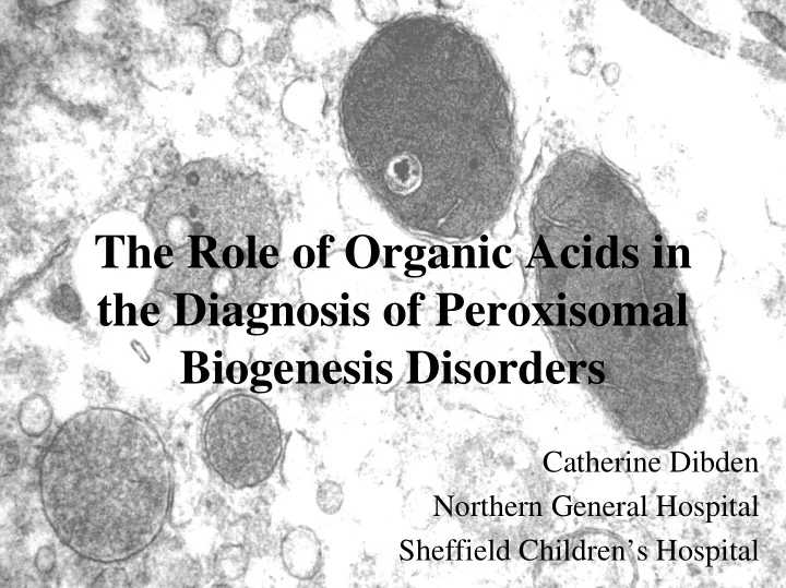

The Role of Organic Acids in the Diagnosis of Peroxisomal Biogenesis Disorders Catherine Dibden Northern General Hospital Sheffield Children’s Hospital
Peroxisomes • Small sub-cellular organelles • Present in all eukaryotic cells • Abundant in tissues actively involved in lipid metabolism – Liver – Kidney – Nervous tissue
Functions of Peroxisomes • Fatty acid β -oxidation • Fatty acid α -oxidation • Ether-phospholipid biosynthesis • H 2 O 2 metabolism • L-pipecolate degradation • Glutaryl-CoA metabolism • Glyoxylate detoxification • Isoprenoid biosynthesis
Peroxisomal Biogenesis • Three steps: – Formation of lipid bilayer – Incorporation of membrane-bound peroxisomal proteins – Import of matrix proteins into the peroxisome • PEX genes encode proteins required for assembly of the peroxisomal membrane and support the import of matrix proteins • Protein products known as peroxins
Peroxisomal Disorders • Single peroxisomal protein deficiencies • Peroxisomal biogenesis disorders: – Rhizomelic Chondrodysplasia Punctata (RCDP) phenotype – Zellweger Spectrum: • Zellweger syndrome (ZS) • Neonatal adrenoleukodystrophy (NALD) • Infantile Refsum disease (IRD) Gould S.J., Raymond G.V., Valle D., 2001. The Peroxisome Biogenesis Disorders, in: C.R. Scriver, A.L. Beaudet, W.S. Sly, D. Valle (Eds.), The Metabolic & Molecular Bases of Inherited Disease. McGraw-Hill, New York
Zellweger syndrome • Presents at birth • Reduction or absence of peroxisomes • Clinical phenotype: – Craniofacial dysmorphism – Hypotonia – Impaired hearing/eye abnormalities – Psychomotor retardation, neonatal seizures – Liver disease – Calcific stippling of epiphyses – Renal cysts • Death usually occurs within 6 months
Zellweger syndrome (2) • Biochemical phenotype: – Plasma: increased very long chain fatty acids (VLCFA), phytanic acid, pipecolic acid, and bile acid intermediates DHCA and THCA – Erythrocytes: reduced plasmalogen synthesis – Fibroblast cultures: reduced dihydroxyacetone phosphate acyltransferase (DHAPAT) activity – Urine: increased pipecolic acid and bile acid intermediates • Diagnosis: abnormal plasma VLCFA levels, confirmed by DHAPAT activity
Problems with Diagnosis of PBD • Plasma VLCFA are not part of routine ‘metabolic screen’ in most metabolic laboratories • Clinicians unfamiliar with rare disorders may not request VLCFA examination • Urine most commonly submitted specimen type for metabolic screening Therefore patients with an undiagnosed PBD may be missed when being screened for a metabolic disease
Role of Organic Acids • GC-MS analysis of urinary organic acids commonly included in the routine ‘metabolic screen’ • Characteristic organic aciduria of PBDs has been reported, showing increased excretion of: – 3,6-epoxydicarboxylic acids (C10, C12, C13, C14) – Odd-chain C7 – C15 dicarboxylic acids – 2-hydroxydecanedioate – Saturated and unsaturated C6 - C10 dicarboxylic acids – C10:C6 and C8:C6 dicarboxylic acid ratios >1 – 4-hydroxyphenyllactic and 4-hydroxyphenylacetic acid Korman et al , 2000. J.Inherit. Metab. Dis. 23: 425 – 428
Aim of Study • To look for the presence of characteristic metabolites and other features of an organic acid profile in patients with a PBD as previously reported • To identify the mass spectra of relevant metabolites for addition to the GC-MS searchable library, to allow routine identification of these metabolites in patient samples.
Methods • Urine from 14 patients with various peroxisomal disorders was examined: – 8 Zellweger Syndrome – 2 Infantile Refsum Disease – 2 X-linked Adrenoleukodystrophy – 1 Pseudo-Zellwegers – 1 Refsum’s Disease • Urine from 20 patients with no specific abnormality on urinary organic acids analysis was also examined • GC-MS analysis of urine organic acids was carried out on all samples
Organic Aciduria of PBD Results:
4-OH phenylacetic acid 3,6-epoxytetradecanedioate Odd-chain dicarboxylic acids Saturated & unsaturated even- 4-OH phenyllactic acid chain dicarboxylic acids C7 C9 2-OH decanedioate C6 C10:1 C10 C8:1 C8 3-OH decanedioate Normal
Mass Spectrum of 3,6-epoxytetradecanedioate
Results • 8/10 patients with PBDs showed increased excretion of: – 3,6-epoxydicarboxylic acids (mostly C14) – 2-hydroxydecanedioate • 3,6-epoxytetradecanedioate: – Specificity: 100% – Sensitivity: 80% • All types of peroxisomal disorders showed elevated levels of odd-chain dicarboxylic acids (mostly C7 and C9)
Results • 7/10 patients with PBDs showed C10:C6 and C8:C6 dicarboxylic acid ratios of >1 – Specificity: 100% – Sensitivity: 70% • Increased levels of 4-hydroxyphenyllactic acid and 4-hydroxyphenylacetic acid were present in patients with a PBD or pseudo-Zellwegers
A few weeks later… • Urine from a 1 month old baby analysed • Clinical details “failure to thrive” • Urine organic acids: – Increased 2-hydroxydecanedioate – Increased 3,6-E14DA • Peroxisomal disorder suspected • Plasma for VLCFA already received, which confirmed diagnosis of a PBD
Conclusions • Urinary organic acids can be a useful indicator to the diagnosis of a PBD • This particular organic acid profile should alert the laboratory to the possibility of a PBD and prompt appropriate investigations, including VLCFAs. • Awareness of the characteristic organic aciduria of a PBD may improve detection of these conditions in an initial metabolic screen
References Gould S.J., Raymond G.V., Valle D., in: C.R. Scriver, A.L. Beaudet, W.S. Sly, D. Valle (Eds.), 2001. The Metabolic & Molecular Bases of Inherited Disease. McGraw-Hill, New York, pp. 3181-3219 Pitt J.J., Poulos A., 1993. Clin Chim Acta. 223: 23-29 Rizzo C., Bertucci P., Federici G., Wanders R.J.A., Sabetta G., Dionisi-Vici C., 2000. J Inherit Metab Dis. 23 (S1): 241. Yamaguchi S., Iga M., Kimura M., Suzuki Y., Shimozawa N., Fukao T., Kondo N., Tazawa Y., Orii T., 2001. J Chrom B. 758: 81-86 www.humpath.com/IMG/jpg/mitochondria_peroxisome_hepatocyte_04-2.jpg Acknowledgements Nigel Manning Sheffield Children’s Hospital Claire Hart Sheffield Children’s Hospital
Recommend
More recommend