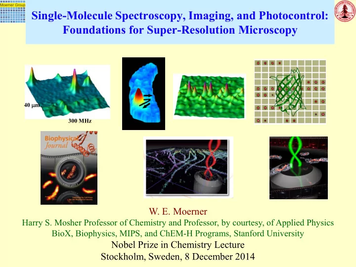

Single-Molecule Spectroscopy, Imaging, and Photocontrol: Foundations for Super-Resolution Microscopy 40 µ m 300 MHz W. E. Moerner Harry S. Mosher Professor of Chemistry and Professor, by courtesy, of Applied Physics BioX, Biophysics, MIPS, and ChEM-H Programs, Stanford University Nobel Prize in Chemistry Lecture Stockholm, Sweden, 8 December 2014
Erwin R. J. A. Schrödinger “…we never experiment with just one electron or atom or (small) molecule. In thought-experiments we sometimes assume that we do; this invariably entails ridiculous consequences…In the first place it is fair to state that we are not experimenting with single particles, any more than we can raise Ichthyosauria in the zoo. “Are There Quantum Jumps? Pt. II” British J. for the Philosophy of Science 3 , 233-242 (1952).
Optical Spectroscopy of Molecules in Solids Spectrum (absorption vs. wavelength, or color) at room T Terrylene in p-Terphenyl Now lets expand the color scale by 25x here, and cool to low T 460 THz=460x10 12 cps 860 THz Frequency=(c/wavelength)
Optical Spectroscopy of Molecules in Solids Spectrum (absorption vs. frequency, or color) at low T (2K) Terrylene in p-Terphenyl Now blow up the scale here 581 nm 569 nm by another 527 THz 516 THz 1000X ! Frequency=(c/wavelength) Data: S. Kummer, … Th. Basché, JChemPhys 1997
Early Steps Toward Single-Molecule Spectroscopy Crucial concepts circa 1985 at IBM Research: •Zero-phonon, purely electronic transitions of fairly flat, rigid molecules in solids at low T become extremely narrow •An inhomogeneous line profile occurs due to tiny variations in local environments •To get homogeneous widths: need photon 5 GHz echoes, … or •Spectral hole-burning: laser-induced changes 506 THz make a dip or “hole” at chosen laser colors What are the ultimate limits to “frequency domain”optical storage by spectral hole-burning? Fundamental Question: Is there a “spectral noise” that results from statistical number fluctuations or the discreteness of individual molecules? – this would define the smallest possible spectral hole that can be detected.
How might statistics appear in spectroscopy? Suppose you have 10 boxes, and throw 50 balls at these boxes with all boxes being equally likely. 6 3 5 6 5 4 5 4 5 7 This is the well-known number fluctuation effect, scaling as √N, that arises from sampling. Key Idea: Now think of the horizontal axis as optical frequency (or wavelength). Each box is a bin of width ∆ν , the homogeneous width of an optical absorption line. Then the resulting spectrum should have a spectral roughness or fine structure scaling as √N, which arises from the discreteness of the individual molecules!
Statistical Fine Structure in an Inhomogeneously Broadened Line WEM and T. P. Carter, Phys. Rev. Lett . 59 , 2705 (1987) pentacene in p-terphenyl crystal, 1.4K •Number of molecules per ∆ν H : N •Fluctuations in N should scale as N •Call this Statistical Fine Structure (SFS) • Detection achieved with FM spectroscopy since it measures ∆α on the 100 MHz scale, or α (upper)- α (lower) SFS arises directly from the discreteness of the individual molecules. The single-molecule limit is within reach! T. P. Carter and WEM, J. Chem. Phys . 89 , 1768 (1988)
Detecting Single-Molecule Absorption PRL 62 , 2535 (May 1989) •Pentacene in crystalline p -terphenyl, 1.8 K, 593 nm •Laser FM absorption spectroscopy with Stark (E-field) or ultrasonic (strain field) secondary modulation •Insensitive to scattering from sample •Limited by laser shot noise (and out-of-focus molecules from relatively thick cleaved crystal) •Challenge: focused laser intensity had to be kept low •Proof-of-principle: single molecules can be optically detected; pentacene/ p -terphenyl is a useful model system FM: G. Bjorklund, Opt. Lett. 5 , 15 (1980) Like FM Radio at 506 THz!
Detecting Single-Molecule Absorption from Emitted Fluorescence •Used the pentacene/ p -terphenyl model system •Detected absorption by measuring emitted fluorescence •Sensitive to scattering from sample, so careful sample growth required – crystal clear sublimed flakes •Limited by Rayleigh and Raman scattering background signals, but produced higher SNR for equal bandwidth PRL 65 , 2716 (November 1990) An application of Laser-Induced Fluorescence (LIF in molecular gas - R.N. Zare, 1968)
Single-Molecule Imaging 0 GHz ≡ 592 nm ≡ 506 THz and Spectroscopy 15000 W. P. Ambrose, WEM, Nature 349 , 225 (1991) 10000 40 microns 5000 Fluorescence (cps) 0 -10.0 0.0 10.0 300 MHz 4000 3000 Spatial scan: SM measures laser spot size with nm probe! 2000 Freq. scan: Second dimension selects one molecule from many in the same focal volume 1000 0 6.0 6.5 7.0 7.5 8.0 GHz Ultralow intensity: Widefield 450 (1.8 mW/cm 2 ) 2D Lifetime-limited 250 Microscopy width: 7.6 MHz Wow! 50 -50 0 50 MHz Güttler,…Wild, ChemPhysLett 217 , 393 (1994)
Some of the Surprises from Single Molecules! Fluorescence excitation spectrum during in repetitive scanning of cw dye laser over (CH 2 -CH 2 -) n 400 MHz at 506 THz: Optically induced spectral shifts! Poisson kinetics observed Molecule spontaneously jumps in frequency space due to nearby host dynamics! W. P. Ambrose, WEM, Nature 349 , 225 (1991) Theory: P. Reilly, J. Skinner, PRL 71 , 4257 (1993) T-dep: A. Zumbusch, M.Orrit, PRL 70 , 3584 (1993) Th. Basché and WEM, Nature 355 , 335 (1992)
Motivations and Impact: Single-Molecule Spectroscopy and Optical Imaging in Complex Systems Remove Ensemble Averaging • Explore heterogeneity : are the various copies identical in behavior, or are they different? • Follow state changes in time, especially in biological processes and complex materials •Test theoretical understanding of stochastic behavior Image/Detect nm-Scale Interactions •Single molecule as a nm-sized reporter and nanometer-sized light source •Distance rulers by FRET, TJ Ha et al. (1996) •Probe local fields in nanophotonic structures • Super-resolution imaging Commercial: Sequence DNA, Imaging •PacBio sequencing with ZMW’s, … …single-photon sources, … •Super-resolution Microscopes
Room Temperature: Milestones of Single- Molecule Detection and Imaging Solution: Fluorescence Correlation Spectroscopy: Magde, Elson, Webb (1972, Correlation 1974); Ehrenberg, Rigler (1974); Pecora (1976); … functions Autocorrelation (FCS) from 1 fluorophore or less in the volume: Rigler, Widengren, BioScience (1990) Solution: Single Multichromophore emitter bursts (phycoerythrin): Peck, Stryer, Glaser bursts Mathies PNAS 86 , 4087 (1989) Single bursts from 1 fluorophore: Shera, Seitzinger, Davis, Keller, Soper, Chem.Phys.Lett. 174 , 553 (1990); Nie, Zare, Science (1994);… Solution and Single antibody with multiple (~80-100) labels: T. Hirschfeld, Appl. surface Opt. 15 , 2695 (1976) Near-Field Imaging a single fluorophore: Betzig and Chicester, Science 262 , 1422 NSOM, SNOM (1993); Ambrose,…, Keller PRL 72 , 160 (1994); Xie and Dunn, Science 265 , 361 (1994) Confocal image Macklin, Trautman, Harris, Brus, Science 272 , 255 (1996); … Widefield, In vitro , myosin on actin: Funatsu,…Yanagida, Nature 374 , 555 (1995). single fluorophore Cell membrane, single-lipid tracking with super-localization, Schmidt, Schütz, …Schindler, PNAS 93 , 2926 (1996).
Detecting Single-Molecule Absorption from Emitted Fluorescence at Room T •Typical organic fluorophore labels are only ~1 nm in size, S 1 S 1 k ISC k ISC fluorescent proteins ~3-4 nm h ν h ν •Light pumps electronic transitions of the molecule •Signal indirectly reports on local nanoenvironment k T k T because only one molecule is pumped and measured, if S 0 S 0 backgrounds are low and molecule emits light efficiently λ /(2NA)~250 nm TMR ~1 nm Cy3 GFP, FPs (~ 3nm x 4 nm)
Single-Molecule Imaging and Tracking Examples – Much to Learn from Isolated Single Molecules! Immune proteins in membrane of a live CHO cell Terrylene molecules in p-terphenyl, Vrljic, Nishimura, McConnell, WEM, Biophys. J. (2002) room T, showing grain boundaries Werley, WEM, J Phys Chem B (2006) Circumferential MreB motions in Caulobacter , Time-lapse stroboscopic tracking, YFP Kim, et al. PNAS (2006) bar 1 µ m
Super-Resolution Microscopy with Single Molecules How can we use single-molecule labels to surpass Abbé’s optical diffraction limit, a fundamental physical effect in the far-field? Sin ingl gle-molecule im imagin aging g + 2 key id ideas eas
Key Idea #1: Super-Localization cinder cone, bar 120x10 9 nm Find the position of the emitter by fitting the shape of the single-molecule image can easily find peak position to much better precision than the width ˆ c center position Fluorescence Intensity 10 2 photons: 20 nm prec. ′ σ ≅ ( ) / ) Abbe N 1 µ m 1 2 3 4 5 6 7 8 9 10 Position (pixels) Summary: WEM, J. Microscopy 246 , 213-220 (2012)
Recommend
More recommend