
Simulation of the Ephaptic Effect in the Cone- Horizontal Cell Synapse of the Retina Carl Gardner, Jeremiah Jones, Steve Baer, & Sharon Crook School of Mathematical & Statistical Sciences Arizona State University
http://webvision.med.utah.edu/
Schematic (Kamermans & Fahrenfort) of horizontal cell dendrite contacting cone pedicle (approx. 1 micron 2 ): simulate 400 nm membranes × 40 nm gap & 10/20 nm openings at side of HC
◮ Experiments show illumination of cone causes hyperpolarization of horizontal cells & increased levels of intracellular cone Ca ◮ Ephaptic hypothesis: specialized geometry of synapse can force currents through high-resistance bottlenecks causing potential drop in extracellular cleft ◮ Cone membrane senses this as depolarization, which increases activation of voltage-sensitive Ca channels ◮ Implies Ca 2 + current is directly modulated by electric potential
Drift-Diffusion (PNP) Model cone pedicle V CP � Φ � � Σ i Ca gap cations Φ � � Σ i V HC � horizontal cell ∂ n i ∂ t + ∇· f i = 0 , i = Ca 2 + , Na + , K + , Cl − , . . . z i = q i f i = z i µ i n i E − D i ∇ n i , j i = q i f i , j = � j i , q e i � E = −∇ φ q i n i , ∇· ( ǫ ∇ φ ) = − i parabolic/elliptic system of PDEs
A Model of the Membrane (similar to Mori-Jerome-Peskin) n i Φ Φ � � �Σ inside Σ i m Φ � � outside �Σ Σ i n i Φ
Poisson-Boltzmann Equation − q i φ � � n i = n bi exp kT �� � − q i φ � � φ � q i n bi exp q 2 i n bi ∇· ( ǫ ∇ φ ) = − ≈ kT kT i i � Debye length l D = ǫ kT / i q 2 i n bi �� � ≈ 1 nm For z ⊥ & near membrane φ zz ≈ φ/ l 2 D 1 − q i φ ± � � φ ≈ φ ± e −| z | / l D , kT e −| z | / l D n i ≈ n ± bi � ∞ q i n i − n + dz ≈ q i l D n + i − n + Set σ + � � � � i = bi bi 0
20 A 15 10 Σ PB 5 M 0 � 3 � 2 � 1 0 1 2 3 u 0 Comparison of nearly exact Poisson-Boltzmann solution for σ i / ( q i n bi l D ) vs. u 0 = q i ( φ 0 − φ b ) / ( kT ) with approximations
Jump conditions for Poisson’s equation [ φ ] ≡ φ + − φ − = V = σ C m n · ∇ φ ] = 0 [ˆ BCs for drift-diffusion equation (Mori-Jerome-Peskin), but we use σ ± n ± i − n ± i = q i l D � � bi ∂ n + ∂σ + i i n · j + = q i l D i − j mi = ˆ ∂ t ∂ t ∂σ − ∂ n − i i n · j − = q i l D i + j mi = − ˆ ∂ t ∂ t � � σ − σ + σ ≡ i = − i i i
Drift-Diffusion Model with Membrane Boundary Conditions ∂ n i ∂ t + ∇· ( z i µ i n i E ) = D i ∇ 2 n i , i = Ca 2 + , Na + , K + , Cl − E = −∇ φ � q i n i , ∇· ( ǫ ∇ φ ) = − bi + σ − CP , HC − σ BCs : n − i i = n − φ − CP , HC = V + , q i l D C m ∂σ − i n · j − � σ − i + j mi , = − ˆ σ = − i ∂ t g Ca ( V CP − E Ca ) ( CP ) j m , Ca = 1 + exp { ( θ − V CP ) /λ m } ( HC ) j hemi = � g i ( V HC − V i ) = g hemi V HC cations V CP , HC ≡ V + CP , HC − φ − CP , HC
cone pedicle V CP � Φ � � Σ i Ca gap cations Φ � � Σ i V HC � horizontal cell n · ∇ φ = 0 BCs at openings are ambient: n i = n bi , ˆ 1. Apply 2D TRBDF2 drift-diffusion code (with SOR for Poisson equation) to cone-horizontal cell problem with model of membrane 2. Investigate relative importance of electrical (ephaptic) [vs. chemical (GABA) or pH] effects
TRBDF2 Numerical Method √ du dt = f ( u , t ) , γ = 2 − 2 u n + γ − γ ∆ t n 2 f n + γ = u n + γ ∆ t n 2 f n ( TR ) γ ( 2 − γ ) u n + γ − ( 1 − γ ) 2 u n + 1 − 1 − γ 1 2 − γ ∆ t n f n + 1 = γ ( 2 − γ ) u n ( BDF2 ) Use Newton’s method if f ( u ) is nonlinear n �Γ n n � 1 TR BDF2
Advantages of TRBDF2 1. One-step (composite) method 2. Second-order accurate & L-stable 3. Easy to adjust ∆ t dynamically C 2 TR TRBDF implant after one timestep 1.5 1 0.5 y 2 4 6 8
Known Biological Parameters Parameter Value Description n b , Ca 10 − 4 , 2 mM intra/extracellular bath density of Ca 2 + n b , Na intra/extracellular bath density of Na + 10, 140 mM n b , K intra/extracellular bath density of K + 150, 2.5 mM n b , Cl intra/extracellular bath density of Cl − 160, 146.5 mM ǫ 80 dielectric coefficient of water N s 20 number of spine heads per cone pedicle A m 0.1 µ m 2 spine head area C m 1 µ F/cm 2 membrane capacitance per area V Ca reversal potential for Ca 2 + 50 mV V Na reversal potential for Na + 50 mV V K reversal potential for K + − 60 mV G hemi 5 nS hemichannel conductance
Known Biological Parameters Parameter Value Description D Ca 0.8 nm 2 /ns diffusivity of Ca 2 + D Na 1.3 nm 2 /ns diffusivity of Na + D K 2 nm 2 /ns diffusivity of K + D Cl 2 nm 2 /ns diffusivity of Cl − 32 nm 2 /(V ns) mobility of Ca 2 + µ Ca 52 nm 2 /(V ns) mobility of Na + µ Na 80 nm 2 /(V ns) µ K mobility of K + mobility of Cl − 80 nm 2 /(V ns) µ Cl
Fitting Parameters in Model for CP Transmembrane I Ca g Ca ( V CP − E Ca ) j m , Ca = 1 + exp { ( θ − V CP ) /λ } Parameter Value Description E Ca cone reversal potential for Ca 2 + 37 mV G Ca 1.5 nS Ca conductance θ off − 33 mV kinetic parameter, bg off (nonlocal) θ on − 40 mV kinetic parameter, bg on (nonlocal) λ 5 mV kinetic parameter Note that g i = G i / ( N s A m ) & I Ca = N s A m j m , Ca da �
Drift-diffusion simulations
Drift-diffusion simulations
Experimental IV curves (Kamermans & Fahrenfort)
10 bkgd off bkgd on 0 bkgd off (exp.) bkgd on (exp.) −10 −20 Current (pA) −30 −40 −50 −60 −70 −80 −90 −70 −60 −50 −40 −30 −20 −10 0 10 Membrane Potential (mV) I Ca vs. V CP shift turning on background illumination
40 d = 10/20 d = 20/40 35 d = 40/80 30 Current Shift (pA) 25 20 15 10 5 0 −70 −60 −50 −40 −30 −20 −10 0 10 Membrane Potential (mV) Ephaptic effect: Shift in I Ca vs. V CP for varying opening widths
Most recent experimental IV curves (Kamermans et al.)
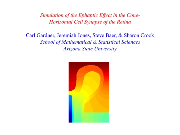
2D Complex Geometry of the Synapse ◮ Replace nonlocal with local BC for bg off/on: add voltage ground ◮ Theoretical argument (Kamermans) that bg off/on produces translation of I Ca curve (without GABA or pH) ◮ Model effects of complex geometry & include bipolar cell ◮ Solve drift-diffusion PDEs inside cells as well as outside ◮ Specify holding potentials U CP , U HC , & U BC as in voltage clamp experiment, & set ground φ = U ref at bottom right corner ◮ Computed potential shows compartment model is not adequate
2D Complex Geometry of Synapse
Finite-Volume (Box) Method & Grid Intracellular + − n Extracellular
U CP = − 15 mV, U BC = − 60 mV, U ref = − 40 mV U HC = − 40/ − 60 mV for bg off/on
New Fitting Parameters in Model for CP Transmembrane I Ca g Ca ( V CP − E Ca ) j m , Ca = 1 + exp { ( θ − V CP ) /λ } Parameter Value Description E Ca cone reversal potential for Ca 2 + 37 mV G Ca 1.5 nS Ca conductance θ 3 mV kinetic parameter (independent of bg) λ 2 mV kinetic parameter
Neutral Bipolar Cell Depolarized Bipolar Cell 0 0 HC/BC = −40/−60 HC/BC = −40/−60 −20 −20 HC/BC = −60/−60 HC/BC = −60/−40 −40 −40 −60 −60 −80 −80 −80 −60 −40 −20 0 −80 −60 −40 −20 0 Hyperpolarized Bipolar Cell Shift Curves 0 100 HC/BC = −40/−60 Neutral −20 HC/BC = −60/−80 Depolarized 50 Hyperpolarized −40 −60 0 −80 −50 −80 −60 −40 −20 0 −80 −60 −40 −20 0 I Ca vs. U CP shift turning on background illumination
Future Work 1. Model effects of GABA & glutamate 2. Model arrays of cones & horizontal cells—homogenize over small spatial scales 3. Multiscale modeling: integrate out shortest time scales in drift-diffusion model to obtain intermediate model, so we can treat time-dependent illuminations of retina
Recommend
More recommend