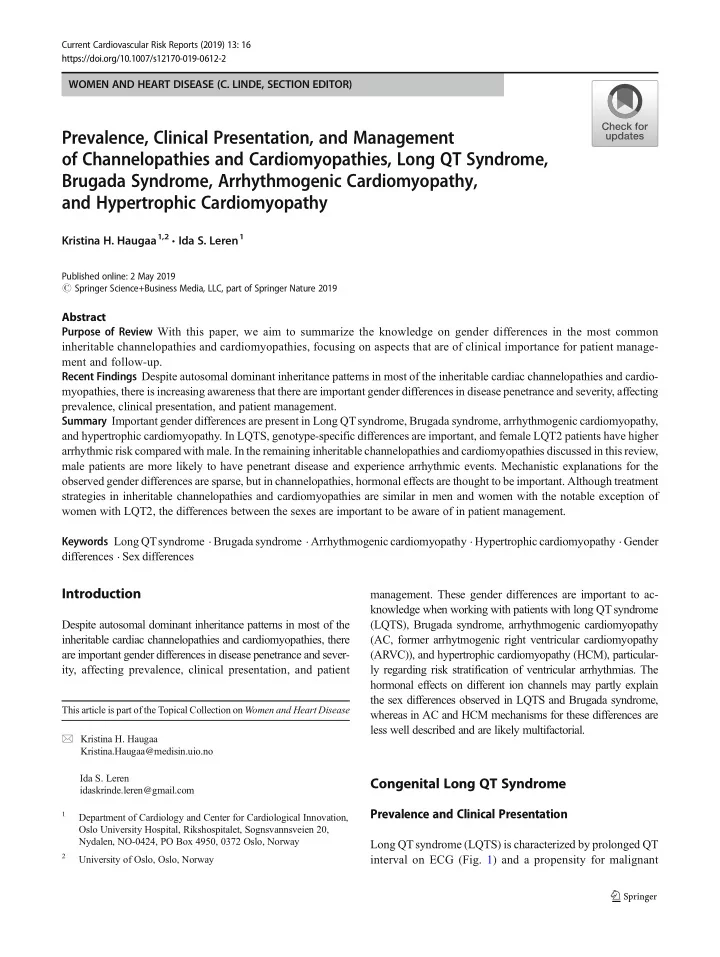

Current Cardiovascular Risk Reports (2019) 13: 16 https://doi.org/10.1007/s12170-019-0612-2 WOMEN AND HEART DISEASE (C. LINDE, SECTION EDITOR) Prevalence, Clinical Presentation, and Management of Channelopathies and Cardiomyopathies, Long QT Syndrome, Brugada Syndrome, Arrhythmogenic Cardiomyopathy, and Hypertrophic Cardiomyopathy Kristina H. Haugaa 1,2 & Ida S. Leren 1 Published online: 2 May 2019 # Springer Science+Business Media, LLC, part of Springer Nature 2019 Abstract Purpose of Review With this paper, we aim to summarize the knowledge on gender differences in the most common inheritable channelopathies and cardiomyopathies, focusing on aspects that are of clinical importance for patient manage- ment and follow-up. Recent Findings Despite autosomal dominant inheritance patterns in most of the inheritable cardiac channelopathies and cardio- myopathies, there is increasing awareness that there are important gender differences in disease penetrance and severity, affecting prevalence, clinical presentation, and patient management. Summary Important gender differences are present in Long QTsyndrome, Brugada syndrome, arrhythmogenic cardiomyopathy, and hypertrophic cardiomyopathy. In LQTS, genotype-specific differences are important, and female LQT2 patients have higher arrhythmic risk compared with male. In the remaining inheritable channelopathies and cardiomyopathies discussed in this review, male patients are more likely to have penetrant disease and experience arrhythmic events. Mechanistic explanations for the observed gender differences are sparse, but in channelopathies, hormonal effects are thought to be important. Although treatment strategies in inheritable channelopathies and cardiomyopathies are similar in men and women with the notable exception of women with LQT2, the differences between the sexes are important to be aware of in patient management. Keywords Long QTsyndrome . Brugada syndrome . Arrhythmogenic cardiomyopathy . Hypertrophic cardiomyopathy . Gender differences . Sex differences Introduction management. These gender differences are important to ac- knowledge when working with patients with long QT syndrome Despite autosomal dominant inheritance patterns in most of the (LQTS), Brugada syndrome, arrhythmogenic cardiomyopathy inheritable cardiac channelopathies and cardiomyopathies, there (AC, former arrhytmogenic right ventricular cardiomyopathy are important gender differences in disease penetrance and sever- (ARVC)), and hypertrophic cardiomyopathy (HCM), particular- ity, affecting prevalence, clinical presentation, and patient ly regarding risk stratification of ventricular arrhythmias. The hormonal effects on different ion channels may partly explain the sex differences observed in LQTS and Brugada syndrome, This article is part of the Topical Collection on Women and Heart Disease whereas in AC and HCM mechanisms for these differences are less well described and are likely multifactorial. * Kristina H. Haugaa Kristina.Haugaa@medisin.uio.no Ida S. Leren Congenital Long QT Syndrome idaskrinde.leren@gmail.com Prevalence and Clinical Presentation 1 Department of Cardiology and Center for Cardiological Innovation, Oslo University Hospital, Rikshospitalet, Sognsvannsveien 20, Nydalen, NO-0424, PO Box 4950, 0372 Oslo, Norway Long QTsyndrome (LQTS) is characterized by prolonged QT 2 interval on ECG (Fig. 1) and a propensity for malignant University of Oslo, Oslo, Norway
Page 2 of 7 Curr Cardiovasc Risk Rep (2019) 13: 16 16 Fig. 1 EGC from a healthy Healthy individual Pa�ent with LQT1 individual (left) and an LQTS patient (right). The QT interval is severely increased in the LQTS patient QTc 380 ms QTc 530 ms 50 mm/s 50 mm/s ventricular arrhythmias, syncope, and sudden cardiac death. Young boys with LQT1 have higher risk of ventricular The QT interval is heart rate – dependent, and therefore the arrhythmias and fatal events compared with girls carrying heart rate – corrected QTc, calculated by, e.g., Bazett ’ s formula the same mutation. However, in puberty, the risk of arrhyth- as QT interval/ √ RR-interval, is used for diagnosis. LQTS is mias is lower in males than females [8]. Therefore, if there the best described ion channelopathy, and sex differences are have been no arrhythmic events until the age of 16, the risk of well known and differ between the specific genetic subtypes. arrhythmias in males decreases, while in females, it remains The QT interval is physiologically longer in women than in the same or increases [8, 9]. men, and previously recommended cutoff values for prolonged QT interval differed between men (QTc > 450 ms) and women (QTc > 460 ms). However, the latest LQT2 guidelines propose a QTc > 480 ms in the absence of second- ary causes for QTc prolongation, to diagnose LQTS for both The sex differences are most obvious in LQT2. Mutations in sexes [1 • ], while a QTc of 460 ms is sufficient to make a the KCNH2 gene cause defect IKr potassium channels, which diagnosis in the presence of unexplained syncope [1 • ]. is the most important potassium channel for repolarization at Genetically determined LQTS has an estimated prevalence rest. Arrhythmias are typically triggered by emotions and loud of 1:2000 [2]. Mutations in genes encoding ion channel sub- noises. Throughout the lifespan, the risk of arrhythmias is units have been found in 60 – 75% of clinically diagnosed higher in females compared with males [8, 10]. Therefore, LQTS patients [3, 4], and in total, 15 genes have been shown women with LQT2 and QTc > 500 ms are considered at higher to be associated with the autosomal dominant form (Romano risk than men with similar findings, and should be evaluated Ward syndrome) and 2 genes with the autosomal recessive for primary preventive ICD [1 • , 11]. Interestingly, in LQT2 subtype (Jervell-Lange Nielsen Syndrome) [5]. Jervell- women, the risk of arrhythmias remains increased also after Lange Nielsen syndrome is characterized by congenital, sen- menopause, suggesting lifelong follow-up and therapy [12]. sorineural deafness in addition to a severe cardiac phenotype In LQT2 women, the risk of arrhythmias increases signif- [6, 7]. icantly in the post-partum period, persisting for the first 9 – Importantly, there are significant sex differences in disease 12 months after delivery [13]. Beta blocker therapy should, penetrance and severity which have to be acknowledged in therefore, be continued during pregnancy and under no cir- patient management. cumstances be reduced in the post-partum period [13]. Management of mothers with LQT2 could also include infor- LQT1 mation that nightly nursing may be handled by the father to ensure adequate rest for the mother, as sleep deprivation is a risk factor for arrhythmic events [14]. A home automatic ex- Mutations in the KCNQ1-gene cause defect IKs potassium ternal defibrillator or a wearable defibrillator may be an option channels with a phenotype of LQT1, which is the most com- in mothers as a bridge during the post-partum period when mon form of LQTS. IKs is most important for normal repo- ICD is not indicated or not preferred although this is not in- larization when heart rate increases, and arrhythmic events are typically triggered by exercise. cluded in current guidelines.
Recommend
More recommend