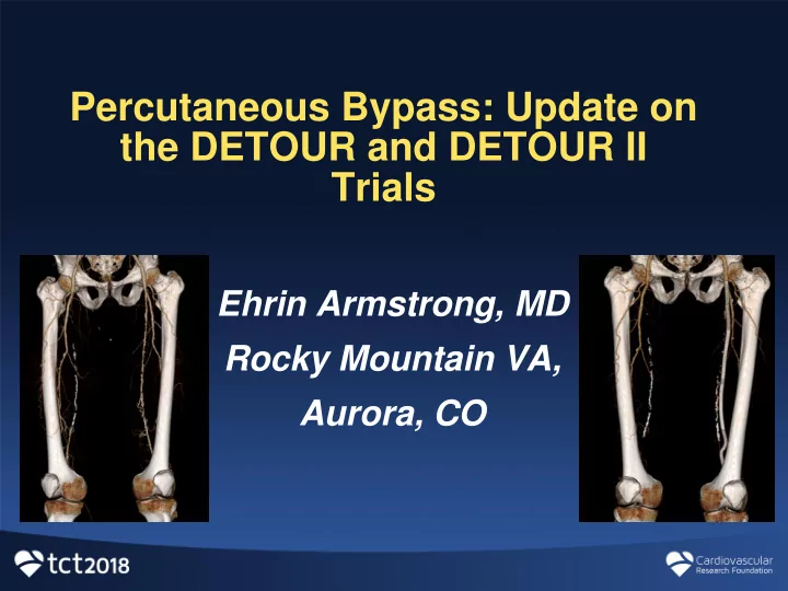

Percutaneous Bypass: Update on the DETOUR and DETOUR II Trials Ehrin Armstrong, MD Rocky Mountain VA, Aurora, CO
Disclosure Statement of Financial Interest I, Ehrin Armstrong, DO NOT have a financial interest/arrangement or affiliation with one or more organizations that could be perceived as a real or apparent conflict of interest in the context of the subject of this presentation.
The Inconvenient Truth About Femoropopliteal Revascularization Despite numerous devices with “long lesion” indications, long, complex SFA lesions do not have an optimal endovascular treatment strategy Simple Lesions Long, Complex Lesions <60% patency at 12M* 80% patency at 12M Endovascular = usually Endovascular = consistently less durable than open first choice for shorter or bypass though 12 months less complex lesions * Composite of the following sources: 1 P100022/S020; 2 P040037/S060; 3 P120002 ; 4 P140010/S037; 5 P070014/S010; 6 P130024; 7 Rocha ‐ Singh, Krishna J., et al. "Patient ‐ level Meta ‐ analysis Of 999 Claudicants Undergoing Primary Femoropopliteal Nitinol Stent Implantation." Catheterization and Cardiovascular Interventions 89.7 (2017): 1250-1256.; 8 P040037; 9 P160025; 10 P120020; 11 P140028; 12 P140002; 13 P160004; 14 P110023
SFA Devices Consistently Demonstrate an Inverse Relationship Between Lesion Length and Durability 100% 12M Primary 90% 59.5% 1-Year Patency Patency 80% 70% 60% Viabahn 25 2 50% Zilver PTX 1 Lutonix 6 40% 30% Everflex 14 20% 10% 0% Lesion Length “Long Lesions” 20.6 cm 30 20 (cm) 10 21.5 26.5 19.2 26.8 12.3 15.9 14.5 19.0 16.5 18.4 28.7 15.2 17.9 18.0 0 1 P100022/S020; 2 P040037/S060; 3 P120002 ; 4 P140010/S037; 5 P070014/S010; 6 P130024; 7 Rocha ‐ Singh, Krishna J., et al. "Patient ‐ level Meta ‐ analysis Of 999 Claudicants Undergoing Primary Femoropopliteal Nitinol Stent Implantation." Catheterization and Cardiovascular Interventions 89.7 (2017): 1250-1256.; 8 P040037; 9 P160025; 10 P120020; 11 P140028; 12 P140002; 13 P160004; 14 P110023 15 Interpolated avg. assuming normal distribution.
The DETOUR Procedure Percutaneous femoropopliteal bypass Surgical principles using an endovascular approach Originates in SFA, travels within the femoral vein, and returns to the popliteal artery Femoral vein becomes pathway for stent graft bypass TORUS ™ Stent Graft DETOUR Crossing Kit
Step 1: Proximal Anastomosis Specialized crossing device and snare create arteriovenous connection above proximal margin of the lesion
Step 2: Distal Anastomosis Specialized crossing device and snare create arteriovenous connection below distal margin of the lesion
Step 3: Graft Deployment Stent graft bypass exits artery, travels within femoral vein, adjacent to occlusion and reenters artery at the site of reconstitution
PQ Bypass Clinical Program CE Mark US Pivotal Proof of Concept (DETOUR I) (DETOUR II) # Subjects 23 81 292 # Centers 1 8 40 IRB-approved, Prospective, Prospective, single- Study Design observational single-arm arm safety and efficacy Follow Up 10 Years 3 Years 3 Years LPI Jul ‘17 Enrollment LPI Ongoing
77 Patients/ 81 Limbs Enrolled DETOUR I • DESIGN: Prospective, single-arm, multi-center clinical evaluation of Follow up at 30D, 3M, 6M, 12M, the DETOUR TM System and 18M, 24M, 36M Procedure for Percutaneous Bypass • INCLUSION CRITERIA: De novo, Primary Efficacy: Primary Safety: CTO, or ISR femoropopliteal Primary Patency MAE at 30D Lesion ≥10 cm; femoral vein at 6M (PSVR ≤ (Death, TLR, diameter ≥10mm or duplicate 2.5) with no TLR Amputation) • Independent Review: Core Lab (DUS, CT, Angio) by Cleveland Clinic; Clinical Events Committee STATUS: CE Mark granted by Syntactx February 2017
Baseline Characteristics N=81 lesions/ 77 Clinical Characteristics N=77 Patients Lesion Characteristics patients Male Gender 83.1% (64/77) Lesion Length 37.1 cm ± 5.5 cm Age, Years 64.1± 7.2 22.2 cm – 47.2 cm Range Diabetes Mellitus 24.7% (19/77) % CTO 96% (78/81) Hypertension 83.1% (64/77) Calcification Hypercholesterolemia 39.0% (30/77) Severe 67.5% (54/80) Moderate 13.8% (11/80) History of CAD or MI 42.9% (33/77) Mild 18.8% (15/80) History of Smoking 87.0% (67/77) TASC II Lesion Type Previous Peripheral 29.9% (23/77) C 56% (45/81) Intervention D 44% (36/81) Vessel Run-off ABI 0.64 ± 0.17 1 8% (6/77) Rutherford 3 92.2% (71/77) 2 29% (22/77) Rutherford 4-5 7.8% (6/77) 3 64% (49/77)
DETOUR I Lesion Distribution by Length Independently adjudicated by Cleveland Clinic Core Laboratory 97.5% 86.4% 71.6% 33.1% > 25 cm > 30 cm > 35 cm > 40 cm Lesion Distribution 450 400 350 Lesion Length (mm) 300 250 200 150 100 50 0 1 3 5 7 9 11 13 15 17 19 21 23 25 27 29 31 33 35 37 39 41 43 45 47 49 51 53 55 57 59 61 63 65 67 69 71 73 75 77 79 81 DETOUR I Lesions (Low to High)
DETOUR I: Efficacy and Safety Through 12 Months Independently adjudicated by Cleveland Clinic Core Laboratory and Syntactx CEC 93.8% 80.0% (75/80) 72.5% Efficacy (64/80) Through 12 (58/80) Months Primary Patency Primary Asst. Patency Secondary Patency PSVR < 2.5 without TLR Revasc of 50%-99% Revasc of 100% occlusion stenoses 30D 12M N=77 Patients/81 Lesions Freedom from DVT 100% (81/81) 97.5% (78/80) Safety Freedom From Death 100% (77/77) 98.7% (76/77) Through 12 Freedom from Amputation 100% (81/81) 100% (80/80) Months Freedom from ALI 98.8% (80/81) 98.8% (79/80) Freedom from TLR 97.6% (79/81) 78.8% (63/80)
Functional Improvement Through 12 Months 90% of patients experienced Rutherford improvement > 2 classes Ankle Brachial Index 100% 90% 80% 12 Months 70% Significant improvement 0.92 ± 0.14 60% at 12M Baseline (p<0.0001) 50% 40% 12M Baseline Significant 30% 0.64 ± 0.17 improvement at 12M 20% (p<0.0001) 10% 0% 5 0 1 2 3 4 Rutherford Becker Clinical Classification 1 8 patients were missing Rutherford- Becker scores at 12M
Trial Update
292 Subjects across 40 centers in US and Europe • DESIGN: Prospective, single-arm, multi-center clinical evaluation of Follow up at 30D, 6M, 12M, the DETOUR TM System and 24M, 36M Procedure for Percutaneous Bypass • INCLUSION CRITERIA: De novo, Primary Safety: Primary Efficacy: CTO, or ISR femoropopliteal MAE at 30D Primary Patency Lesion ≥15 cm; femoral vein (Death, TLR, at 12M (PSVR diameter ≥10mm or duplicate Amputation, ≤2.5) with no TLR DVT) • Independent Review: Core Lab (DUS, CT, Angio) by Cleveland Clinic; Clinical Events Committee STATUS: Enrollment ongoing by Syntactx
Case Review: Rocky Mountain VA • 72 year-old male • 35 cm TASC D lesion • Rutherford 3 • History of smoking • BMI of 30
Case Review: Pre Procedural Imaging SFA Popliteal Venogram
Case Review: Proximal Crossing Alignment of Crossing Device Marker Crossing Device Needle from Artery into Vein – Wire Advanced into Snare Band in SFA; Snare Expanded in FV
Case Review: Distal Crossing Crossing Device Docked with Snare in Wire advancement into Vein; Needle firing in orthogonal view popliteal artery
Case Review: Pre and Post Angiogram Pre Post
Case Review: Pre and Post Venogram Pre Post
Conclusions ❑ Safety outcomes from DETOUR I demonstrate percutaneous bypass has a promising safety profile with 100% freedom from amputation, 98.8% freedom from acute limb ischemia and 98.7% freedom from death at 12 months ❑ Excellent durability in long, challenging, occlusive lesions (Cleveland Clinic Core Lab Adjudicated Patency) ❑ 72.5% Primary Patency, 78% Primary Assisted Patency, 93.8% Secondary Patency ❑ 12-Month safety and durability outcomes in DETOUR I demonstrate fully- percutaneous bypass is a promising endovascular alternative for complex femoropopliteal disease DETOUR II IDE designed to build upon extant body of clinical evidence in even longer, more challenging lesions
Recommend
More recommend