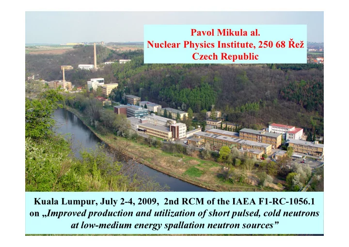

Pavol Mikula al. Nuclear Physics Institute, 250 68 Řež Czech Republic Kuala Lumpur, July 2-4, 2009, 2nd RCM of the IAEA F1-RC-1056.1 on „ Improved production and utilization of short pulsed, cold neutrons at low-medium energy spallation neutron sources”
Our main task within the contract 14198 : Development and optimization of a curved wide- wavelength band monochromator based on strongly cylindrically bent perfect Si-slabs in a sandwich for microfocusing small-angle neutron scattering (mfSANS) device The main research task on the side of NPI Řež was preparation of crystal slabs for a sandwich type monochromator for a newly designed compact SANS instrument as proposed by Hokkaido University (prof. M. Furusaka) which would permit us to demonstrate a new type of inexpensive instrument which can be designed and realized for operation at different neutron wavelengths. Our laboratory has to provide know-how in the design and optimization performance of the required wide wavelength band focusing monochromators. Furthermore, our laboratory has to provide Si- perfect crystals of a special cut for cylindrical bending.
Neutron production Reactor LVR 15, NRI Řež, CZ • reactor power 10 MW • thermal flux in the core 1.5 10 18 ns -1 m -2 • beam tube 1 10 13 ns -1 m -2 • fuel enrichment 36% 235 U • tank type • light water moderated and cooled
Experimental facilities installed at the reactor LVR-15 in Řež NBCT strain scanner II SANS powder diffractometer multipurpose diffractometer NDP Radiative capture powder strain scanner I diffractometer 0 1 2 m
Neutronový difractometr SPN 100 Sample Monochromator drum Detector unit
What we have done? For Hokkaido University Two sets (2x30) of the perfect silicon crystal slabs of a special cut with the lattice planes (111) at the angle 67.5 deg have been prepared. The dimensions of the slabs are: 120x20x0.5 mm 3 (length x width x thickness). Therefore, such a cut of the crystal slabs permits us to use the bent focusing monochromator in the so called fully asymmetric diffraction geometry when employing it just at the Bragg angle of 67.5 deg. Two sets (1x30 and 1x40) of the crystal slabs of the same orientation which will be used for mechanical tests (minimum curvature, optimum number of slabs in the sandwich) have been prepared. For HMI Berlin Two horizontally and vertically focusing monochromators and three sets of Si crystals with the main face parallel to 311, 331 and 400 lattice planes. For KAERI Daejeon One horizontally focusing monochromators and 17 Si crystal slabs of different cut. For JINR Dubna One horizontally focusing monochromators and Si(220) and Si(111) crystal slabs.
Collaborative works Horizontally and vertically focusing Horizontally focusing monochromator monochromator manufactured for HMI. manufactured for KAERI different 2 pieces, with Si(311) and Si(400) thicknesses of the Si(111) crystals and planes, figure of merit increased 10x. different asymmetric geometries.
Monte Carlo simulations KAERI Daejeon CIAE Beijing NECSA, South Africa JINR Dubna – Kurchatov Inst. Moscow
Future plans and collaboration We are developing a long term collaboration with KAERI Daejeon. Within 2009/10 we have to prepare two new horizontally focusing monochromators including Si(111) slabs of different thicknesses Construction of the horizontally and vertically focusing mono-chromator for CIAE Beijing for China Advanced Research Reactor Construction of the horizontally and vertically focusing monochromator for Mirrotron Budapest manufacturing whole stress diffractometer for Mianyang institute in Sichuan Construction of the horizontally focusing monochromator for BARC Mumbai (bending device ready, crystals in preparation) Construction of the horizontally focusing monochromator for Malaysian Nuclear Agency Monte Carlo simulations for KAERI Daejeon After clarifying some problems related to the lower efficiency of the present monochromator, new sets of crystals will be prepared for HU.
SANS instrumentation Double-crystal system – slit geometry Double-bent-crystal high-resolution Ultra high-resolution Bonse-Hart SANS camera SANS camera
At home Reconstruction of the high resolution small - angle neutron scattering double crystal diffractometer: •New collimator with the saphire filter •New monochromator shielding •Improved shielding between individual instruments •Improved sample environment Reconstruction of the multipurpose neutron diffractometer: •New detector arm (Huber) • PSD detector •New monochromator shielding •Improved shielding between individual instruments
SANS Instrumentation size range SANS technique 1 nm .. 100 nm Collimator instruments 10 nm .. 1 µ m DC diffractometers with bent crystals 1 µ m .. 10 µ m DC diffractometers with perfect crystals (Bonse-Hart) � Developed experimental technique � Advanced data evaluation method & software − multiple scattering − Non-linear data fitting of a single model to multiple data sets
Double-Crystal SANS Diffractometer DN-2 beam tube with collimator beam shutter Instrument Parameters Monochromator bent perfect crystal Si 111 symmetric geometry bent Si 220 maximum 5x25 mm 2 Sample Analyzer bent perfect crystal Si 111 bent Si 111 fully asymmetric geometry 1-dimensional 3 He PSD Detector resolution ~ 1 mm Pb Wavelength λ = 2.1 A, ∆λ / λ < 0.01 bent Si 111 analyzer PE + B (asymmetric cut) 5 ⋅ 10 3 n s -1 cm -2 / R M = 300 m Neutron flux samples 4 ⋅ 10 4 n s -1 cm -2 / R M = 35 m x = ( sin(2 R ) + L ) ∆ θ ∆θ D A D S 1 ⋅ 10 -4 A -1 / R M = 300 m Q-resolution 1 ⋅ 10 -3 A -1 / R M = 35 m ∆ x D steel rods L D 10 -1 A -1 Vertical Q-resolution Total Q-range 2 10 -4 ÷ 2 10 -2 A -1 ∆θ S position sensitive detector diffraction planes 111
SANS-Diffractometer in NPI
Small-angle neutron scattering Study of porosity in plasma-sprayed ceramics SANS data for various resolutions Size distribution of pores in and sample thickness fitted to a plasma-sprayed Al 2 O 3 single model. 140 10000 z=5 mm as sprayed o C T=1730 15 120 o C 1300 1000 -1 ] o C 1520 100 -2 µ m z=2.5 mm 10 100 o C z d Σ /d θ 1730 80 D(R) [10 10 5 60 empty beam 1 40 0 0.1 1 0.1 20 0.01 0 1 10 100 0.01 0.1 1 -1 ] Q x [ µ m R [ µ m] Collimator instruments Bonse-Hart
The most recent results Experimental powder diffraction test at a small take-off angle
Experimental test PSD detector Bent perfect crystal monochromator α -Fe sample 2 mm diameter 40 mm width Monochromator take-off angle 30 o . No collimators were used.
Diffraction geometries Ψ = 22 o , t=4mm Ψ = 29.5 o , t=3 mm Ψ = 35.26 o , t=4mm B B P P C-monochromator C-monochromator Ψ Ψ Ψ = 0 o , t=4 mm Ψ = 0 o , t=3x1.3 mm 2θ=30 deg 2θ=30 deg Ψ = 0 o , t=1.3 mm Asymmetric transmission PSD detector PSD detector geometry, OBC BP BP C-monochromator C-monochromator Ψ=0 deg Ψ 2θ=30 deg 2θ=30 deg PSD detector PSD detector Symmetric reflection geometry Spatial resolution of the PSD – 2 mm 1 channel = 0.009 o
MC simulations (output beam expansiom) Si 111, chi=35.26, 1/R opt =0.33 200 200 200 Ge 111, chi=29.5, 1/R opt =0.31 Si 111, chi=29.5, 1/R opt =0.29 Intensity [n/s] 150 150 150 100 100 100 200 Si 111, chi=0, 1/R opt =0.11 50 50 50 150 0 0 0 -30-20-10 0 10 20 30 -30-20-10 0 10 20 30 -30-20-10 0 10 20 30 x [mm] x [mm] x [mm] 100 Si 111, chi=-48.53 1/R opt =-0.25 Si 111, chi=48.53 1/R opt =0.40 200 200 200 Si 111, chi=22, 1/R opt =0.26 50 150 150 150 Intensity [n/s] 0 100 100 100 0 -30-20-10 0 10 20 30 x [mm] 50 50 50 0 0 0 -30-20-10 0 10 20 30 -30-20-10 0 10 20 30 -30-20-10 0 10 20 30 x [mm] x [mm] x [mm]
MC simulations (output beam compression) Si 111, chi=-35.26, 1/R opt =-0.155 Ge 111, chi=-29.5, 1/R opt =-0.11 Si 111, chi=-29.5, 1/R opt =-0.11 200 200 200 Intensity [n/s] 150 150 150 200 100 100 100 Si 111, chi=0, 1/R opt =0.11 50 50 50 150 0 0 0 -30 -20 -10 0 10 20 30 -30 -20 -10 0 10 20 30 -30 -20 -10 0 10 20 30 x [mm] x [mm] 100 x [mm] Si 111, chi=-48.53 1/R opt =-0.24 Si 111, chi=-22, 1/R opt =-0.05 200 200 50 150 150 Intensity [n/s] 0 100 100 0 -30-20-10 0 10 20 30 x [mm] 50 50 0 0 -30 -20 -10 0 10 20 30 -30 -20 -10 0 10 20 30 x [mm] x [mm]
Experimental results: Asymmetric transmission geometry (OBC) si(111) Ψ =35.26 deg si(111) Ψ =22 deg FWHM FWHM Output beam compression 75 Output beam compression 750 Peak height Peak height 200x40x4 mmm 200*30*4 920 FWHM / channel numbers 62 FWHM / channel numbers 70 700 900 Peak height 60 880 Peak height 65 650 860 58 60 600 840 55 550 56 820 800 50 500 54 40 60 80 100 120 140 140 160 180 200 220 240 Bending / µ m Bending / µ m Ψ =29.5 deg FWHM 60 800 Output beam compression Peak height 200x30x3 mm FWHM / channel numbers 750 Peak height 55 700 50 650 45 600 100 120 140 160 180 200 Bending / µ m
Recommend
More recommend