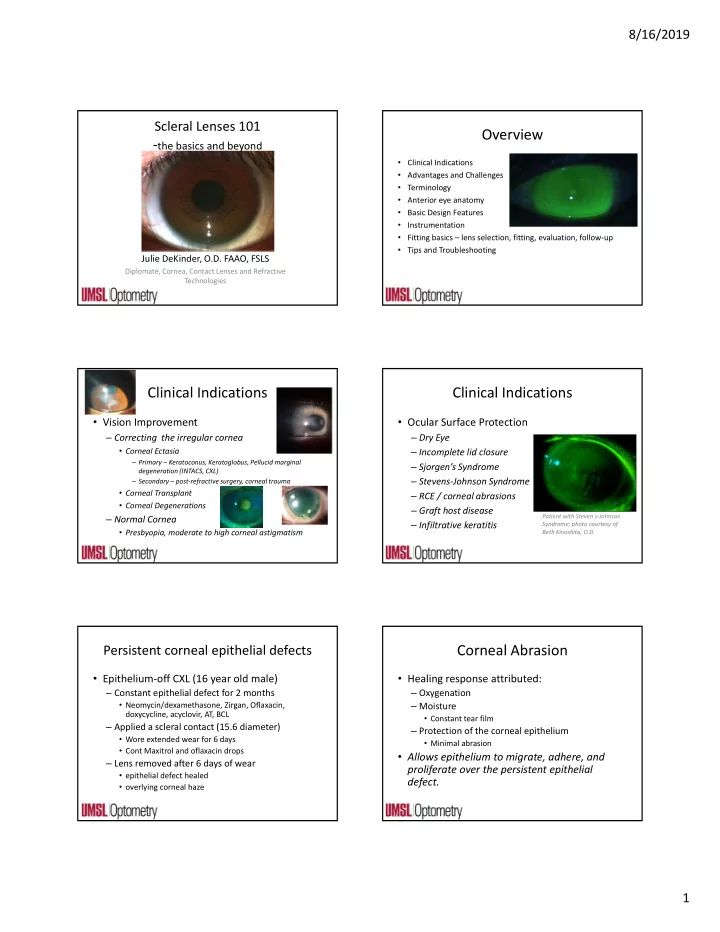

8/16/2019 Scleral Lenses 101 Overview ‐ the basics and beyond • Clinical Indications • Advantages and Challenges • Terminology • Anterior eye anatomy • Basic Design Features • Instrumentation • Fitting basics – lens selection, fitting, evaluation, follow‐up • Tips and Troubleshooting Julie DeKinder, O.D. FAAO, FSLS Diplomate, Cornea, Contact Lenses and Refractive Technologies Clinical Indications Clinical Indications • Vision Improvement • Ocular Surface Protection – Correcting the irregular cornea – Dry Eye • Corneal Ectasia – Incomplete lid closure – Primary – Keratoconus, Keratoglobus, Pellucid marginal – Sjorgen’s Syndrome degeneration (INTACS, CXL) – Stevens‐Johnson Syndrome – Secondary – post‐refractive surgery, corneal trauma • Corneal Transplant – RCE / corneal abrasions • Corneal Degenerations – Graft host disease – Normal Cornea Patient with Steven s‐Johnson – Infiltrative keratitis Syndrome; photo courtesy of • Presbyopia, moderate to high corneal astigmatism Beth Kinoshita, O.D. Corneal Abrasion Persistent corneal epithelial defects • Epithelium‐off CXL (16 year old male) • Healing response attributed: – Constant epithelial defect for 2 months – Oxygenation • Neomycin/dexamethasone, Zirgan, Oflaxacin, – Moisture doxycycline, acyclovir, AT, BCL • Constant tear film – Applied a scleral contact (15.6 diameter) – Protection of the corneal epithelium • Wore extended wear for 6 days • Minimal abrasion • Cont Maxitrol and oflaxacin drops • Allows epithelium to migrate, adhere, and – Lens removed after 6 days of wear proliferate over the persistent epithelial • epithelial defect healed defect. • overlying corneal haze 1
8/16/2019 Advantages of Scleral GPs vs Corneal Clinical Indications GP • Cosmetic/Sports • Centration – Hand‐painted scleral lenses – Fitting a “regular” part of the eye • Lens Retention – Ptosis – Water sports – Minimal chance of inferior standoff • Comfort – Reduced lid interaction; no corneal interaction • Lens failure in other designs • Vision – Masking severe corneal irregularity Challenges associated with scleral Terminology lenses • Handling • Classification – Difficult I and R (initially) – Apprehensive patients – Corneo‐scleral 12.9mm to 13.5mm • Fitting – Semi‐Scleral 13.6 mm to 14.9mm – Subtle fit indications – Mini‐Scleral 15.0mm to 18.00mm – Increased chair time – Full‐Scleral 18.1mm to 24+ • Physiology – Dk/L – Oxygen must diffuse over great distance – Long‐term effects of scleral lens wear are unknown Anatomy and Shape of the Anterior Terminology Ocular Surface • Maximum scleral lens size for normal eye: 24mm • Scleral Shape Study Scleral Lens Education Society June 2013 A guide to scleral lens fitting 2.0 www.sclerallens.org Assuming 12mm cornea diameter – maximum physical diameter of a scleral lens ~24 mm 2
8/16/2019 Anatomy and Shape of the Anterior Anatomy and Shape of the Anterior Ocular Surface Ocular Surface • Clinical Consequences • Corneal Toricity does not typically extend to – Temporal‐Inferior decentration of scleral sclera lenses • Inferior decentration • The ocular surface beyond the cornea is – Weight/gravity nonrotationally symmetrical – Eyelid pressure • Temporal – Asymmetrical – Flatter nasal elevation – The entire nasal portion typically flatter compared • Conjunctival Prolapse to the rest Basic Design Features Basic Design Features • Spherical Design • Spherical Design • Concentric symmetrical (spherical) scleral lens • Concentric symmetrical (spherical) scleral lens • Non‐toric back surface • Non‐toric back surface – Optic Zone – Transition Zone • Centermost zone • Optics/Lens power • Mid‐periphery or limbal zone Same optics rules apply as – Anterior surface • Creates the sagittal height corneal GP • Back surface • Can be reserve geometry – Ideally mimics corneal shape • Completely vaults limbus • Completely vaults cornea Basic Design Features Basic Design Features • Spherical Design • Concentric symmetrical (spherical) scleral lens • Non‐toric back surface Optical/Transition Zone Base Curve – Landing Zone PC 1 PC2 • Area of the lens that rests on anterior ocular surface Landing Zone • Scleral zone or haptic PC3 Example Parameters: PC4 • Alignment to provide even BC: 7.50 pressure distribution is key PC1: 7.85 (if reverse geometry 6.89) PC2: 9.00 PC3: 12.25 PC4: 14.00 3
8/16/2019 Basic Design Features Basic Design Features • Toric Lens Designs • Toric Lens Designs – Back Toric Haptics – Front Surface Toric ‐ • Landing zone is made toric to improve lens fit • Anterior surface front toric optics to improve vision • Does not interfere with central zone of scleral lens • Located on the front surface of the central optical zone • Better ocular health • Indicated when residual cylinder over‐refraction is – Fewer areas of localized pressure found – Decreased bubble formation • Needs stabilization – Longer wearing time and better patient comfort – Dynamic stabilization zones or prism ballast • More frequently needed in larger diameter sclerals – LARS Basic Design Features • Toric Lens Designs – Bitoric both anterior optics and back toric haptics • Front surface toric optical power • Back surface toric periphery • No need for lens stabilization Basic Design Features Basic Design Features • Multifocal Scleral lens design • Multifocal Scleral lens design – Simultaneous Multifocal Lens Design • Aspheric or concentric • Center Near and Center Distance Designs – Can adjust near powers – Can adjust zone size • Not all scleral lens designs have a MF option http://www.aldenoptical.com/products/soft‐specialty/zen‐multifocal/zen‐ multifocal/ 4
8/16/2019 Basic Design Features Fitting Basics • Lens Material • Hydra‐PEG – High(est) Dk lens material; plasma or hydra‐PEG – Polyethylene glycol (PEG) – base polymer • Considerably thicker when compared to corneal GP • Covalently bonded to the lens surface – 250 microns to 500 microns • Creates a wetting ocular surface, increases surface • Optimum Extreme, Menicon Z wettability, increases lubricity, decreases protein and lipid deposits, improves TBUT. • Increasing Oxygen transmissibility – 1. high Dk material (Dk > 125) Monthly conditioning – 2. minimal tear clearance behind the lens (<200) Cleaning and solution to restore the disinfecting coating – 3. Reduced center thickness of the lens (<.250) Fitting Basics Fitting Basics • Completely vault the cornea and limbus while How can I vault a steep cornea with a flat lens? aligning to the bulbar conjunctiva BC much flatter than “K” Very steep cornea Fitting Basics Fitting Basics • 1. Diameter • 2. Clearance • 1. Diameter • 3. Landing Zone Fit – Choose a Fitting Set • 4. Lens Edge • Direct vs Indirect control – Laboratory warranty/exchange policy • 5. Asymmetrical Back Surface Design – Overall Diameter • Some trial sets are toric back surface • Larger – more clearance needed, ectasias • 6. Lens Power • Smaller – easier to handle, less clearance 5
8/16/2019 Fitting Basics Fitting Basics • 1. Diameter • 2. Clearance – HVID – Minimum of ~100 microns • <12mm – Typically aim for 200‐300 microns after settling – Start with a 16.0 mm or smaller lens – Maximum of 600 (if desired) • >12mm – Start with a 16.0 mm or larger lens – Base Curve Determination – Diameter of the optical zone and the transition • Select an initial base curve that is flatter than the flat k zone chosen roughly 0.2mm larger than the value corneal diameter • Use 14 mm chord OCT, measure sagittal depth Fitting Basics Fitting Basics Estimate Corneal Clearance • Evaluate overall corneal chamber appearance – Diffuse beam, low mag, medium illumination Lens – Observe centration, areas of bearing, tear lens appearance, look for bubbles Tear Lens Cornea Fitting Basics Fitting Basics Look for continuity of the tear lens… • Evaluate central clearance Acceptable clearance: Too little clearance: *Compare lens thickness to tear lens thickness and estimate central clearance in microns Christopher Gilmaritn, OD 6
8/16/2019 Fitting Basics Fitting Basics Look for continuity of the tear lens… Evaluate the Limbal Clearance… Fitting Basics Fitting Basics • Change lens base curve/sagittal depth until • UMSL Study: desired central clearance is reached – No significant settling after 4 hours of wear – Most settling within the 1 st hour – Considerations: • All scleral lenses will settle over a period of hours • Expect ~ 90 to 150 microns settling – Large Diameter Scleral settle ~90 microns, slower • Aim for 150 to 300 microns after settling – Mini Scleral ~130 microns, faster • Build‐in settling time into fitting session ~30 min Fitting Basics Fitting Basics • Evaluate remaining corneal chamber • Anterior Segment OCT – Optic Section – Sweep limbus to limbus noting tear lens thickness – Looking for tears in optic section beyond the limbus and should increase in thickness toward the central cornea ** Adequate limbal clearance is critical for an acceptable fit and good tear exchange** 7
Recommend
More recommend