mRNA purification through fragmentation Distinct 28S ribosomal - PowerPoint PPT Presentation
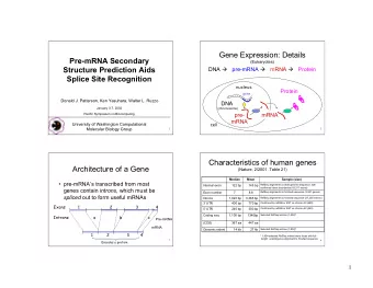
mRNA purification through fragmentation Distinct 28S ribosomal Distinct 18S subunit (or prok. 23S): ribosomal 28S ideally 2X size of 18S subunit (or prok. 16S) 18S No well defined peaks between Flat baseline Marker throughout
mRNA purification through fragmentation
Distinct 28S ribosomal Distinct 18S subunit (or prok. 23S): ribosomal 28S ideally 2X size of 18S subunit (or prok. 16S) 18S No well defined peaks between Flat baseline Marker throughout ribosomal peaks electropherogram ~100 bp Small peaks are sometimes present after the marker at 24 – 29 Marker seconds. These are represented by 5S and 5.8S subunits, tRNAs, and small RNA fragments about 100bp. These are especially noted when using phenol and trizol extraction methods. They can be removed by treating total RNA through Qiagen columns which removes small RNAs.
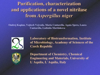
Total RNA with images like this are borderline. Re-extraction should be seriously considered. Decrease in ribosomal peak intensities The 28S subunit often degrades first Increase in intensity of smaller digested RNA fragments Baseline between and to the left of ribosomal peaks becomes jagged
Decrease in ribosomal peak intensities ~100 bp marker Digested RNA Combination of 5S, 5.8S, tRNAs, and an increase in digested RNAs
~100 bp
Relies on base pairing between poly-A tail and oligo dT beads
Fragmentation Buffer: Mg2+ and Heat Stop Solution: EDTA
Used for concentrating and de-salting DNA or RNA in an aqueous solution Components: ◦ Salt (NaAcO): RNA has a negatively charge backbone NaAcO breaks up to Na+ and [CH3COO]- Salt ions neutralize the charge on the nucleic acids, the molecules become less hydrophillic and less soluable ◦ EtOH Easier for Na+ to interact with the phosphate backbone of the nucleitides in EtOH than in water because EtOH is less polar Image from: http://openwetware.org/wiki/Image:DNA_EtOH_precipitation_small_pellet.jpg
1 st Strand Synthesis 2 nd Strand Synthesis End Repair 3’ Adenylation Ligation Same as DN DNA Proto tocol Gel Extraction PCR Amplification
Wear gloves and use sterile technique at all times. Reserve a set of pipettes for RNA work Use sterile RNase ‐ free filter pipette tips to prevent cross ‐ contamination Use disposable plasticware that is certified to be RNase ‐ free. All reagents should be prepared from RNase ‐ free components Store RNA samples by freezing. Avoid extended pauses in the protocol until the RNA is in the form of double ‐ stranded (ds) DNA Use a RNase/DNAse decontamination solution to decontaminate work surfaces and equipment prior to starting this protocol
Recommend


More recommend
Explore More Topics
Stay informed with curated content and fresh updates.