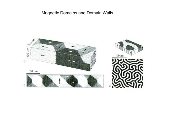

Magnetic Domains and Domain Walls
Bitter-Technique (ferromagnetic colloids on surfaces) Stress induced domains in NiFe single crystal
Magneto-optics Magneto-optical Kerr Effect
Magneto-optical Kerr Effect
Kerr microscopes
Domains in Fe single crystal
Transmission electron microscopy Lorentz microscopy
Scanning electron microscopy with polarization analysis (SEMPA) SiFe single crystal Co single crystal (0001)
Magnetic Force Microscopy Image: Border of a 30 µm wide bit track of 300 kfrpi (89 nm bit length)
Photo electron emission microscopy Electrons
Excited oscillation modes 16x32 µm 2 10 nm Py/Cu I I I h h h 1 1 1 2 2 2 H H Oscillating field , 1GHz, 0.2mT A XMCD ~ M . ~ cos PhD-thesis of A. Krasyuk
SP-STM and magnetic contrast E E e - Tip Sample ρ ρ parallel dI ( U ) n ( 0 ) n ( eU ) n ( 0 ) n ( eU ) n n ( eU ) m m ( eU ) cos ( M , M ) T S T S T S T S T S dU E E Tip Sample ρ ρ antiparallel
Tip and sample preparation T. Duden and E. Bauer, M. R. S. Proc.,287 (1997). Co Co Au(111) Au(111) W(110) W(110) [110] W tips - flashing 2000 K W(110) evaporation - RT 10 ML Au Au 4 – 16 ML Co Co
Out-of-plane magnetic contrast 4 ML Co/Au/W T = 5 K I = 1.5 nA U = 0.3V T = 5 K I = 1.5 nA U = 0.4V Fe [001] Fe _ [110] Mo Mo 100 nm Fe Fe __ 100 nm dI __ dI 100 nm dU dU (500 500) nm 2 (500 500) nm 2 Fe Fe height [Å] height [Å] Mo(110) Mo(110) lateral displacement [nm] lateral displacement [nm]
Domain wall width in the ML Fe nanowires 1.2 ± 0.2 nm 150 4 ML Co/Au/W T = 5 K I = 1.5 nA U = 1 V 100 Z[mV] 50 0 Fe -50 0 2 4 6 8 10 12 14 X[nm] Topography Mo Fe 40nm 14nm 40nm __ dI (200 200) nm 2 [ 001 ] dU __ (70 70) nm 2 dI dU [ 1 1 0 ]
Domain wall width in Fe on W(110) Domain wall Width: w ML = 6 Å Energy: e ML =15.2 meV/atom row Exchange stiffness A = 3.6 pJ/m (8.2 meV/atom) Anisotropy constant K = 41 MJ/m 3 (4.0 meV/atom) M. Pratzer, H.J. Elmers, M. Bode, O. Pietzsch, A. Kubetzka, and R. Wiesendanger, Phys. Rev. Lett. 87, 127201 (2001).
A. Hubert and R. Schäfer Springer 1998
Recommend
More recommend