
Klinik für Gastroenterologie und Hepatologie
Etiologies of Upper GI Bleeding • Duodenal ulcerations 27% • Gastric ulcerations 24% • Varices 19% • Gastroduodenal erosions 13% • Reflux esophagitis 10% • Mallory-Weiss lesions 7% • Tumores 3% • Angiodysplasia 1% • not identifiable 6% Ell, DMW 1995 Klinik für Gastroenterologie und Hepatologie
Esophageal Varices - Epidemiology - Risk of development in liver cirrhosis: 30-40% with compensated cirrhosis 60 % with decompensated cirrhosis New onset of esophageal varices in liver cirrhosis 5-10%/year 2 ° : 1/3 of luminal 1 ° : collaps on insufflation 3 ° : >50% of luminal diameter diameter Klinik für Gastroenterologie und Hepatologie
Esophageal Varices - Epidemiology - Total bleeding risk of esophageal varices 25-50% Factors determining risk of hemorrhage HPVG >12mmHg Large varices Child-Pugh stage MELD score Alcohol consumption Mortality after hemorrhage (up to 50% in 6 weeks) 70% re-bleeding within first year without secondary prophylaxis García - Pagán , Sem Respir Crit Care Med 2012 Klinik für Gastroenterologie und Hepatologie
Esophageal Varices - Therapeutic Scenarios - • Primary prevention • Acute variceal bleeding • Prevention of recurrent bleeding Klinik für Gastroenterologie und Hepatologie
Variceal Bleeding - Primary Prevention- Non-selective betablockers Band ligation Small varices without risk ± no factors Small varices with red yes no spots or CHILD C Medium or large varices Either betablockers or band ligation Use hepatic venous pressure gradient (HPVG) for estimation of indication/prognosis (if available) De Franchis, J Hepatol. 2010 (Baveno V Consensus Workshop) Klinik für Gastroenterologie und Hepatologie
Esophageal Variceal Bleeding - Preendoscopic therapy - - Venous access (multiple large catheters) - Volume resuscitation - ICU treatment, stabilization - Blood transfusions (hemoglobin cut-off 7g/dl) - Pharmacotherapy: terlipressin (on suspicion of variceal bleeding) Terlipressin Placebo Active VB (endoscopy) 17% 28% Recurent bleeding (12h) 12% 26% Mortality (20d) 20% 42% Levacher, Lancet 1995 De Franchis, Dig Liver Dis. 2004 Klinik für Gastroenterologie und Hepatologie
Esophageal Variceal Bleeding - Endoscopic standard therapy - Band ligation superior to sclerotherapy (early and long term results) Therapy Primary Early Complications (+pharmacoth.) failure recurrence Band ligation 4% 5% 14% Sclerotherapy 15% 9% 28% Villanueva, J Hepatol 2006 Klinik für Gastroenterologie und Hepatologie
Esophageal Variceal Bleeding - Endoscopic standard therapy - Klinik für Gastroenterologie und Hepatologie
Esophageal Variceal Bleeding - Endoscopic standard therapy - Klinik für Gastroenterologie und Hepatologie
Esophageal Variceal Bleeding - TIPS - Transiugular Intrahepatic Portosystemic Shunt Hepatic vein TIPS Portal vein Klinik für Gastroenterologie und Hepatologie
Esophageal Variceal Bleeding - TIPS - TIPS in high-risk patients after EBL High risk: Child B + active bleeding Child C (all pts) Failure of therapy Early TIPS: Recurrent bleeding 1year mortality Garcia-Pagan, N Engl J Med. 2010 Problem: TIPS in salvage situation – death in >50% Klinik für Gastroenterologie und Hepatologie
Survey 01 Klinik für Gastroenterologie und Hepatologie
Esophageal Variceal Bleeding - Treatment Failure - Failure to control bleeding Baveno IV - Time frame 120 hours - Fresh hematemesis ≥2 hours after treatment / endoscopic intervention - >3g/dl drop in hemoglobin (no transfusions) - Death - Adjusted blood transfusion requirement index (ABRI)≥0.75 Baveno V - Time frame 120 hours - Fresh hematemesis / NG tube aspiration ≥2 hours after treatment / endoscopic intervention - >3g/dl drop in hemoglobin (no transfusions) - Hypovolemic shock or death De Franchis, J Hepatol 2005 De Franchis, J Hepatol. 2010 Klinik für Gastroenterologie und Hepatologie
Esophageal Variceal Bleeding - Treatment Failure - Balloon tamponade Sengstaken – Blakemore - Tube Limited time frame (<24 hours, if possible) Frequent decompression necessary to avoid esophageal necrosis High complication rate – aspiration / regurgitation / perforation Panes, Dig Dis Sci 1988 Klinik für Gastroenterologie und Hepatologie
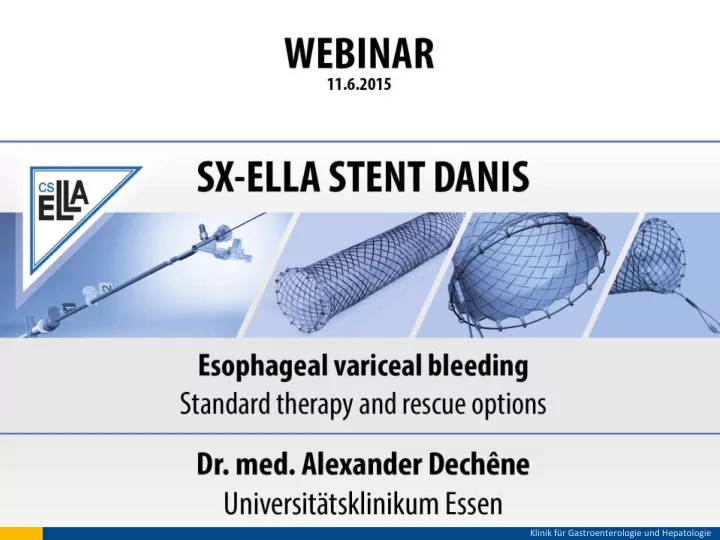
Esophageal Variceal Bleeding - Treatment Failure - Self-expanding metal stent (SEMS) SX Ella Stent DANIS Device properties: - fully covered metal stent - flares on both ends - retrieval lassos on both ends - delivery system with positioning balloon Work principle: - distension of esophageal wall - compression of esophageal varices - termination of hemorrhage Klinik für Gastroenterologie und Hepatologie
SX Ella Stent Danis - System Demonstration - Klinik für Gastroenterologie und Hepatologie
SX Ella Stent Danis - Recommendations for Placement - SEMS placement possible without endoscopic guidance Confirm esophageal bleeding source whenever possible Use a guidewire (guide wire included) when possible Adhere strictly to implantation manual Endoscopic and/or radiographic guidance during stent deployment possible Klinik für Gastroenterologie und Hepatologie
SX Ella Stent Danis - Follow-up care - Confirm proper stent placement by endoscopy as soon as possible Check stent position after 24h (by X-ray or endoscopy) or in signs of bleeding After stent placement, stabilize pt. and evaluate TIPS indication Remove stent after a week, longer indwelling time often possible Remove stent urgently on suspicion of airway compression Klinik für Gastroenterologie und Hepatologie
Survey 02 Klinik für Gastroenterologie und Hepatologie
SX Ella Stent Danis - Extraction - Klinik für Gastroenterologie und Hepatologie
SX Ella Stent Danis - Clinical Case - Klinik für Gastroenterologie und Hepatologie
SX Ella Stent Danis - Published Data- Pilot study 11/02-05/05 143 episodes of esophageal variceal bleeding 15 refractory bleedings + 3 pts. with balloon compression + 2 pts. without primary endoscopic therapy Three stent designs (diameter 18-25mm, length 95-140mm) Stent indwelling time 1 – 14d Hubmann, Endoscopy 2006 Klinik für Gastroenterologie und Hepatologie
SX Ella Stent Danis - Published Data- Immediate hemostasis in all patients Stent removal in 18/20 pts (n=2 fatal liver failure) No primary complications with explant Complementary n 60-day- treatment mortality TIPS 5 (28%) n = 0 Surgical shunt 5 (28%) n = 0 Band ligation 5 (28%) n = 1 Medical 2 (11%) n = 1 Hubmann, Endoscopy 2006 Klinik für Gastroenterologie und Hepatologie
SX Ella Stent Danis - Published Data- Extended cohort (2008) 01/03-08/06 34 SEMS in eosphageal variceal bleeding (all SX-Ella) Implantation without complications, n=7 distal dislocations (partial) Stent indwelling time1 – 14d, median 5d Complementary n No recurrent bleeding with indwelling stent treatment TIPS 8 (24%) No recurrent bleeding 30d after SEMS removal Surgical shunt 5 (15%) 60-day mortality n=10 (29%) Band ligation 11 (32%) Medical ? Zehetner, Surg Endoscopy 2006 Klinik für Gastroenterologie und Hepatologie
SX Ella Stent Danis - Published Data- 2010 10 SEMS in esophageal variceal hemorrhage (all SX-Ella) n=5 failure of primary endoscopic treatment n=3 unsuccessful balloon compression n=2 eophageal perforation on balloon compression 9/10 successful implantation (1/10: dysfunction of positioning balloon) 7/9 immediate hemostasis (2/9: bleeding source distally to esophagus) Wright, Gastrointest Endoscopy 2010 Klinik für Gastroenterologie und Hepatologie
SX Ella Stent Danis - Published Data- Follow up: 42d-survival 50% 4/10 sustained hemostasis (>60d), 2xTIPS 1/10 early recurrence (30d), successful EBL+TIPS 2/10 death by exsanguination (distal bleeding) 1/10 death by multi-organ failure with indwelling stent 2/10 death by multi-organ failure after stent removal Wright, Gastrointest Endoscopy 2010 Klinik für Gastroenterologie und Hepatologie
SX Ella Stent Danis - Published Data from Essen, Germany- 8 pts. with esophageal variceal hemorrhage (08/07-08/09) 5 male, 3 female, median age 62 years (1 pt. treated twice with SEMS) Acute bleeding episodes, refractory to pharmacological treatment and EVL Klinik für Gastroenterologie und Hepatologie
SX Ella Stent Danis - Published Data from Essen, Germany- 9/9: EV-hemorrhage and SEMS implant 9/9: immediate hemostasis 6/9 only pharmacologic treatment 3/9 Intervention directed at to lower portal pressure lowering portal pressure 1/6 Death after 5d No recurrent bleeding with indwelling SEMS 3/3 Stent removal after 5/6 SEMS removal after 10 ± 1,5 d (8-11d) 10 ± 3,6 d (6-12) 1/5 Emergency SEMS removal (bronchus compression) 3/3 SEMS removal after intervention and stabilization 4/5 SEMS removal after Death 13 days after SEMS removal stabilization Dechêne, Digestion 2012 without further bleeding Klinik für Gastroenterologie und Hepatologie
Recommend
More recommend