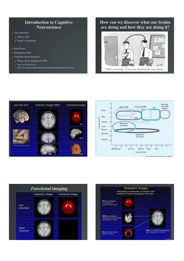

Introduction to Cognitive How can we discover what our brains Neuroscience are doing and how they are doing it? � Tom Johnstone � Office: 1S25 � Email: i.t.johnstone � Brain basics � Imaging the brain � Automatic threat detection � Please check blackboard AND: http://www.beclab.org.uk � http://www.cognitiveneurosciencearena.com/whatiscognitiveneuroscience.asp � Churchland and Sejnowski, 1988 Functional imaging
PET: Positron Emission Tomography PET: Positron Emission Tomography � Uses radioactive tracers introduced into the blood � The tracers bind to some molecule of interest in the brain � Amount and source of emitted radiation tells us about amount and location of molecule of interest � Used to image blood, oxygen, neurotransmitters (e.g. Dopamine) � � Slow (minutes - hours) � Measuring dopamine neuromodulation in the thalamus: Using [F-18]fallypride PET to PET Tracers study dopamine release during a spatial attention task Some PET Radiocompounds and Their Biomedical Applications: 15O-oxygen Oxygen metabolism � 15O-carbon monoxide Blood volume � 15O-carbon dioxide Blood flow � 13N-ammonia Blood flow � 18F-fluorodeoxyglucose Glucose metabolism � 18F-fluoromisonidazole Hypoxic cell tracer � 11C-SCH23390 Dopamine DI receptor � 11C-flumazenil Benzodiazepine receptor � (Adapted from www.austin.unimelb.edu.au/dept/nmpet/pet/detail/radionuc.html) Christian et al., 2005: http://dx.doi.org/10.1016/j.neuroimage.2005.11.052 EEG: Electroencephalography EEG � Measures electrical field on scalp � Electric field changes with neural activity in the brain � EEG can detect fast changes to the electrical field (e.g. < 50 milliseconds) � � Cannot tell us exactly where the source(s) of the electrical field changes are
EEG: Electroencephalography http://www.mrc-cbu.cam.ac.uk/research/eeg/eeg_intro.html MRI: Magnetic Resonance Imaging MRI: Magnetic Resonance Imaging � Uses an extremely powerful electromagnet to detect brain structure and function � Magnetic field: 3 Tesla: approx. 60,000 x Earth's magnetic field � Functional MRI (fMRI): based on magnetic properties of haemoglobin with and without attached oxygen � 3T Siemens Trio MRI scanner MRI: Magnetic Resonance Imaging fMRI � Detects changes in oxygenated blood concentration due to changes in blood flow
fMRI FMRI: Typical experiment � Reasonable time resolution (a few seconds), but not � Repeatedly scan participants (one complete brain as fast as EEG (or many neural processes!) � scan every 2-3 seconds) while they perform a task � Good 3D spatial resolution: can identify source of � Two or more conditions (e.g. Angry versus Happy MRI signal to within a few mm faces, easy versus difficult task, warm heat versus painful heat) � � Non-invasive, but still certain restrictions (no metal or pacemakers!) and LOUD!!! � Continue until enough repetitions of each task condition have been collected (approx. � Relative measure: must always have a within-subject 20/condition) � control condition (no purely between-subjects designs) � � Typically acquire 30-60 minutes of data FMRI: Emotional pictures
Questions to ask yourself whenever FMRI: Emotional pictures reading a Cognitive Neuroscience research paper: � What can it really tell us about function? Does it tell us more than a behavioural experiment? (and is it worth the cost of $3 million per scanner, then $700 per hour)? � Would it be as convincing without the pretty pictures of brains? � Are the identified regions of the brain meaningful functional units? More generally, is the brain modular? � Is it good science? What can it really tell us about Are the identified regions of the brain function? meaningful functional units? � Coltheart (2004) poses the question: � Most cognitive neuroscience techniques have a spatial resolution of half a centimetre or more. This � "Has cognitive neuroscience, or if not might it ever (in is a large region in comparison to the size of neurons principle, or even in practice) successfully use(d) data or networks and nucleii of neurons. from cognitive neuroimaging ... to adjudicate between competing information-processing models of some � Do the “blobs” of activation (neuroimaging), lesions cognitive system)?" (p. 21). (neuropsychology) or virtual lesions (TMS) encompass more than one possible functional unit? Coltheart, M. (2004) Brain imaging, connectionism and cognitive neuropsychology. Cognitive Neuropsychology, 2, 21-25. Coltheart, M. (2006). What has functional neuroimaging told us about the mind (so far)? Cortex, 42(3), 323-31. Example: Patient SM: bilateral Example: LeDoux’s model of fear amygdala damage � Adolphs et al. (‘94) � SM 30 years, normal intelligence � She had trouble: � recognizing facial expressions of fear � generating the facial expression of fear � drawing an expression of fear
OMG!!! My Brain Hurts!! Amygdala inputs
Recommend
More recommend