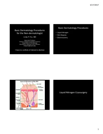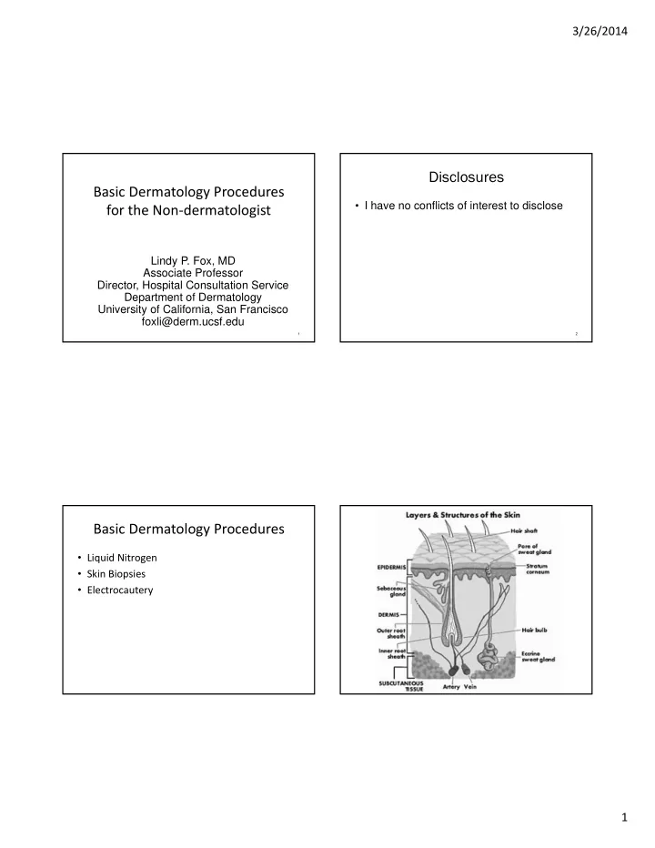
3/26/2014 Disclosures Basic Dermatology Procedures • I have no conflicts of interest to disclose for the Non ‐ dermatologist Lindy P. Fox, MD Associate Professor Director, Hospital Consultation Service Department of Dermatology University of California, San Francisco foxli@derm.ucsf.edu 1 2 Basic Dermatology Procedures • Liquid Nitrogen • Skin Biopsies • Electrocautery 1
3/26/2014 Liquid Nitrogen Cryosurgery • Indications – Benign, premalignant, in situ malignant lesions Liquid Nitrogen Cryosurgery • Objective – Selective tissue necrosis • Reactions predictable – Crust, bulla, exudate, edema, sloughing • Post procedure hypopigmentation – Melanocytes are more sensitive to freezing than keratinocytes Liquid Nitrogen Cryosurgery Principles • ‐ 196°C ( − 320.8°F) • Temperatures of − 25°C to − 50°C ( − 13°F to − 58°F) within 30 seconds with spray or probe • Benign lesions: − 20°C to − 30°C ( − 4°F to − 22°F) • Malignant lesions: − 40°C ( − 40°F) to − 50°C. • Rapid cooling intracellular ice crystals • Slow thawing tissue damage • Duration of THAW (not freeze) time is most important factor in determining success From: Bolognia, Jorizzo, and Schaffer. Dermatology 3 rd ed. Elsevier 2012 Am Fam Physician. 2004 May 15;69(10):2365 ‐ 2372 2
3/26/2014 Liquid Nitrogen Cryosurgery • Fast freeze, slow thaw cycles – Times vary per condition (longer for deeper lesion) – One cycle for benign, premalignant – Two cycles for warts, malignant (not commonly done) • Lateral spread of freeze (indicates depth of freeze) – Benign lesions 1 ‐ 2mm beyond margins – AKs ‐ 2 ‐ 3mm beyond margins – Malignant ‐ 3 ‐ 5+mm beyond margins (not commonly done) Liquid Nitrogen Cryosurgery Technique • Hold spray gun 1 ‐ 1.5cm away from target • Freeze until ice field fills the margin • Maintain the spray for the appropriate time BEYOND initial time of ice field formation • If more than one cycle required, allow for complete thawing before beginning next cycle 3
3/26/2014 Cryosurgery for Common Warts Cryosurgery for Planar Warts • Freeze time 20 ‐ 60 seconds • Margin ‐ 2 ‐ 3mm • May consider • Thaw 30 ‐ 45 seconds cotton tipped • TWO cycles better than one applicator • Repeat every 3 ‐ 4 weeks technique • Average # of warts cleared= 40% • Average # of treatments to clear warts = 12 – ONE YEAR! Ring Wart Bullae http://www.dermnet.com 4
3/26/2014 Cryosurgery for Actinic Keratoses Cryosurgery for Seborrheic Keratoses • One freeze ‐ thaw cycle • Freeze ‐ thaw cycle depends on thickness • margin ‐ 2 ‐ 3mm • Thin/flat ‐ freeze 5 ‐ 10s • Freeze time • Large/thick ‐ freeze >10s, – AK 5 ‐ 7s may need second cycle – Actinic cheilitis 10 ‐ 20s www.dermquest.com Cryosurgery for SCC in situ * Cryosurgery for Lentigines • One 30 second freeze • Quick 3 ‐ 4s freeze Or • Avoid overfreeing • Two 20 second freezes – Risk of hypopigmentation • Close follow up *ED+C still preferred treatment option 5
3/26/2014 Skin Biopsy • Procedure itself is easy • Knowing when and where to biopsy much Skin Biopsies more difficult • Pathologist can only comment on the tissue provided (not what’s left on patient) • Potential pitfalls in technique Skin Biopsy Types Curettage with Biopsy • Curettage • Samples epidermis only • Clinically benign lesions involving the epidermis • Snip/scissors – Verrucae (warts), seborrheic keratoses, actinic • Shave biopsy keratoses • Saucerization • Send pathology at same time as treating the • Punch lesion • Incisional • Limitations – Limited to the epidermis • Excisional ( in toto ) – Fragmented tissue 6
3/26/2014 Snip/Scissors Biopsy • Pedunculated lesions • Benign growths – Acrochordons (skin tags) – Filiform warts – Pedunculated nevi • Hold like pencil • Draw pressure under the lesion (epidermis) From: Bolognia, Jorizzo, and Schaffer. Dermatology 3 rd ed. Elsevier 2012 Shave Biopsy • Samples epidermis and papillary (superficial) dermis • Ideal for elevated lesions involving the epidermis and superficial dermis – Inflammatory dermatoses of epidermis, superficial dermis (psoriasis, eczema, CTCL, lichen planus) – Nevi, benign adnexal tumors • If very thin attachment to skin (stalk) don’t need anesthesia – Diagnosis of basal cell or squamous cell carcinoma • Use iris or Gradle scissors – Diagnosis of lentigo maligna (MIS) • May require hemostasis with aluminum chloride, electrodesiccation From: Bolognia, Jorizzo, and Schaffer. Dermatology 3 rd ed. Elsevier 2012 7
3/26/2014 www.hovesskinclinic.co.uk • Be sure to get below simple hyperkeratosis and upper dermis • Palms, soles, hyperkeratotic lesions • Require hemostasis with aluminum chloride, electrodesiccation From: Bolognia, Jorizzo, and Schaffer. Dermatology 3 rd ed. Elsevier 2012 Onsurg.com Am Fam Physician. 2011 Nov 1;84(9):995 ‐ 1002 Saucerization Biopsy • Deeper biopsy with intentional deeper placement of the blade • Samples epidermis and superficial and deep dermis • Advantage – Histologic examination of the entire circumference of the lesion with adequate depth to assess invasion • Ideal for – Inflammatory dermatoses with dermal infiltrate • Intention is to get to deep dermis – Atypical pigmented lesions (to r/o melanoma) • Requires hemostasis with aluminum chloride, electrodesiccation – Keratoacanthoma/SCC From: Bolognia, Jorizzo, and Schaffer. Dermatology 3 rd ed. Elsevier 2012 8
3/26/2014 Punch Biopsy Punch Biopsy • Samples epidermis, dermis and superficial subcutaneous fat • Varying barrel sizes ‐ 2mm ‐ 8mm • Ideal for – Inflammatory dermatoses with deep dermal infiltrate (lupus) – Infiltrative diseases (amyloid, sarcoid, lymphoma cutis) – Blistering diseases (pemphigus, pemphigoid) – Depressed lesions (scleroderma) • Limitations • Stabilize skin around punch with free hand – Only samples portion of larger lesion • Twist with firm downward pressure in one direction – Requires suture (>3mm) • Gently lift tissue with forceps at edge of epidermis (do not crush) – Not ideal for subcutaneous lesions • If plug not elevating, angle scissors downward to base • Try to make sure there is some fat at the base of the sample Slide courtesy of Wilson Liao, MD Incisional Biopsy • Samples epidermis, dermis, subcutaneous fat • Removes wedge from center or edge of lesion • Ideal for – Large tumors – Subtle diseases of connective tissue – Diseases of the fat (panniculitis) – Diseases of the fascia From: Bolognia, Jorizzo, and Schaffer. Dermatology 3 rd ed. Elsevier 2012 9
3/26/2014 Excisional Biopsy Skin Biopsies ‐ Potential Pitfalls • Samples epidermis, dermis, subcutaneous fat • Crush artifact • Intended to be definitive treatment • Leaving part of tissue in punch tool • Ideal for • Multiple specimens, mislabeling – Suspected invasive melanoma Crush Artifact Failure to Deliver • Leaving part of the biopsy in the punch tool Biopsy Slide courtesy of Jeff North, MD Slide courtesy of Jeff North, MDc 10
3/26/2014 Multiple Biopsy Specimens Shave Biopsy Tray • Critically important to have an established protocol/routine to ensure the correct biopsy goes in the correct bottle C A B Slide courtesy of Jeff North, MD Punch Biopsy Tray How to biopsy a specific lesion Lesion Type of biopsy Papulosquamous (eczema, Shave or saucerization biopsy psoriasis) r/o melanoma Saucerization or excisional biopsy Blister Punch biopsy at the edge for H+E and DIF Wart, seborrheic keratosis, Shave biopsy or curettage actinic keratosis Scalp (alopecia) Punch biopsy from hair containing region adjacent to alopecia, request transverse sections 11
3/26/2014 Where to Biopsy Direct Immunofluorescence Lesion Location of biopsy • Location of the biopsy depends on diagnosis Tumor Thickest portion, avoid necrotic tissue • Vasculitis ‐ lesional skin from an early lesion Blister Edge of the lesion, include about 2mm • Lupus of blister edge; send for H+E and DIF – DLE/SCLE Lesional skin Ulceration/necrotic Edge of ulcer or necrosis plus adjacent – SLE ‐ Lesional, uninvolved lesion skin can be positive as wel Generalized Characteristic lesion of recent onset (+/ ‐ polymorphic eruption more developed lesion) • Blistering Small vessel vasculitis Characteristic lesion of recent onset – Peri ‐ lesional (palpable purpura) (ideally <24 hours old) Adapted from: Bolognia, Jorizzo, and Schaffer. Dermatology 3 rd ed. Elsevier 2012 Slide courtesy of Jeff North, MD DIF in Pemphigoid and Pemphigus DIF ‐ peri ‐ lesional • Eclipsing the edge of new blister • Being too far from a blister can cause false negative DIF Slide courtesy of Jeff North, MD Slide courtesy of Jeff North, MD Photo courtesy of Kari Connolly, MD 12
3/26/2014 DIF in Other Immunobullous Disease • Dermatitis herpetiformis • Up to 1 cm away from lesion Electrosurgery • Don’t overlap the clinical lesion • Higher risk for loss of epidermis and destruction of Ig by the neutrophilic inflammatory infiltrate • Serology: anti ‐ transglutaminase and anti ‐ endomysium antibodies also helpful Slide courtesy of Jeff North, MD Electrosurgery Electrosurgery • Electrodesiccation • Electrodesiccation – Superficial tissue destruction – Superficial tissue destruction • Electrocoagulation • Electrocoagulation – Deep tissue destruction – Deep tissue destruction • Electrosection • Electrosection – Cutting – Cutting 13
Recommend
More recommend
Unleash a World of Digital Possibilities—Browse, Share, and Explore Content Without Boundaries