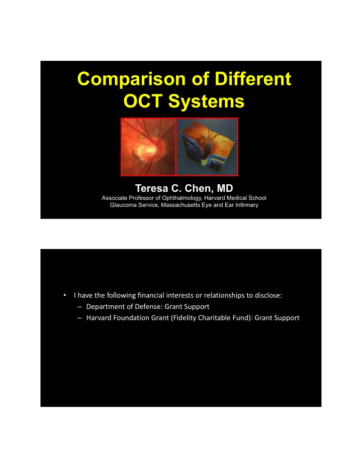
Comparison of Different OCT Systems Teresa C. Chen, MD Associate - PDF document
Comparison of Different OCT Systems Teresa C. Chen, MD Associate Professor of Ophthalmology, Harvard Medical School Glaucoma Service, Massachusetts Eye and Ear Infirmary I have the following financial interests or relationships to
Comparison of Different OCT Systems Teresa C. Chen, MD Associate Professor of Ophthalmology, Harvard Medical School Glaucoma Service, Massachusetts Eye and Ear Infirmary • I have the following financial interests or relationships to disclose: – Department of Defense: Grant Support – Harvard Foundation Grant (Fidelity Charitable Fund): Grant Support
Outline Purpose Review the literature on the use of SD‐OCT to help diagnose glaucoma Methods 2006 to 2018 All commercially available SD‐OCT machines Results RNFL, optic nerve, macula not lamina Conclusions no OCTA
Outline Purpose Methods PubMed and Cochrane Library Databases February 2006 to April 2018 Results Conclusions Outline Purpose Methods PubMed and Cochrane Library Databases February 2006 to April 2018 Results Conclusions
Outline Purpose Methods PubMed and Cochrane Library Databases February 2006 to April 2018 Results Lin SC, Singh K, Jampel HD, Hodapp EA, Conclusions Smith SD, Francis BA, Dueker DK, Fechtner RD, Samples JS, Schuman JS, Minckler DS. Optic Nerve Head and RNFL Analysis. Ophthalmology 2007;114:1937‐1949. Outline Purpose Methods PubMed and Cochrane Library Databases February 2006 to April 2018 Results Lin SC, Singh K, Jampel HD, Hodapp EA, Conclusions Smith SD, Francis BA, Dueker DK, Fechtner RD, Samples JS, Schuman JS, Minckler DS. Optic Nerve Head and RNFL Analysis. Ophthalmology 2007;114:1937‐1949.
Outline Purpose Methods PubMed and Cochrane Library Databases February 2006 to April 2018 Results Chen TC, Hoguet A, Junk A, Nouri‐Mahdavi K, Conclusions Radhakrishnan S, Takusagawa H, Chen PP. Spectral Domain OCT: Helping the Clinician Diagnose Glaucoma. Ophthalmology 2018;125:1817‐1827. How Time Domain OCT Works Reference Mirror SLD Light Source Beam Huang D, Swanson EA, Splitter Lin CP, Schuman JS, Stinson WG, Chang W, Hee MR, Flotte T, Gregory K, Puliafito CA, Fujimoto JG. Optical Coherence Tomography. Photo Detector A-line Science, 1991.
How Spectral Domain OCT Works Reference Mirror SLD Light Source Beam Splitter Spectrometer Fourier transform Johannes de Boer PhD (MGH) “Ultra High Speed Optical Coherence Tomography” Video-Rate SD-OCT White BR, Pierce MC, Nassif N, Cense B, Park BH, Chen TC, de Boer JF…Imaging Using Ultra-High- Speed Spectral Domain Optical Doppler Tomography. Optics Express 2003; 11 (25): 3490-7. American Glaucoma Society Sarasota, Florida, 2004
SDOCT (3D = Video-Rate) 3D Spectral Domain OCT - 2003 imaging the eye for the first time in 3D (video-rate) wide field! in real time White BR, Pierce MC, Nassif N, Cense B, Park BH, Chen TC, de Boer JF…Imaging Using Ultra-High-Speed Spectral Domain Optical Doppler Tomography. Optics Express 2003; 11 (25): 3490-7. Nassif N, Cense B, Park BH, Yun SH, Chen TC, Bouma BE, Tearney GJ, de Boer JF. In vivo Human Retinal Imaging by Ultrahigh-Speed Spectral Domain OCT. Opt Lett 2004;29(5):480-482. Nassif N, Cense B, Park BH, Pierce M, Yun SH, Bouma BE, Tearney GJ, Chen TC, de Boer JF. In vivo High-resolution Video-Rate Spectral Domain OCT of the Human Retina and Optic Nerve. Opt Express 2004;12(3):367-376. Cense B, Nassif N, Chen TC, Pierce MC, Yun SH, Park BH, Bouma BE, Tearney GJ, de Boer JF. Ultrahigh-resolution High-speed Retinal Imaging Using Spectral Domain OCT. Opt Express 2004;12(11):2435-2447. 708 articles Inclusion criteria: - SD-OCT was the technology Outline - RNFL, optic nerve, macula - original research - SD-OCT & glaucoma diagnosis - adult subjects - at least 125 patients Exclusion criteria: Purpose - reproducibility - progression - level III evidence Methods PubMed and Cochrane Library Databases February 2006 to April 2018 Results Chen TC, Hoguet A, Junk A, Nouri‐Mahdavi K, Conclusions Radhakrishnan S, Takusagawa H, Chen PP. Spectral Domain OCT: Helping the Clinician Diagnose Glaucoma. Ophthalmology 2018;125:1817‐1827.
708 articles Inclusion criteria: - original research Outline - SD-OCT was the technology - RNFL, optic nerve, macula - SD-OCT & glaucoma diagnosis - adult subjects - at least 125 patients Exclusion criteria: Purpose - reproducibility - progression - level III evidence Methods PubMed and Cochrane Library Databases February 2006 to April 2018 Results Chen TC, Hoguet A, Junk A, Nouri‐Mahdavi K, Conclusions Radhakrishnan S, Takusagawa H, Chen PP. Spectral Domain OCT: Helping the Clinician Diagnose Glaucoma. Ophthalmology 2018;125:1817‐1827. 708 articles Inclusion criteria: - original research Outline - SD-OCT was the technology - RNFL, optic nerve, macula - SD-OCT & glaucoma diagnosis - adult subjects - at least 125 patients Exclusion criteria: Purpose - reproducibility - progression - level III evidence Methods PubMed and Cochrane Library Databases February 2006 to April 2018 Results Chen TC, Hoguet A, Junk A, Nouri‐Mahdavi K, Conclusions Radhakrishnan S, Takusagawa H, Chen PP. Spectral Domain OCT: Helping the Clinician Diagnose Glaucoma. Ophthalmology 2018;125:1817‐1827.
708 articles Inclusion criteria: - original research Outline - SD-OCT was the technology - RNFL, optic nerve, macula - SD-OCT & glaucoma diagnosis - adult subjects - at least 125 patients Exclusion criteria: Purpose - reproducibility - progression - level III evidence Methods PubMed and Cochrane Library Databases February 2006 to April 2018 Results Chen TC, Hoguet A, Junk A, Nouri‐Mahdavi K, Conclusions Radhakrishnan S, Takusagawa H, Chen PP. Spectral Domain OCT: Helping the Clinician Diagnose Glaucoma. Ophthalmology Inclusion & exclusion criteria yielded: 2018;125:1817‐1827. - 2 level I articles - 57 level II articles Outline Many different SD‐OCT machines Purpose Methods Results Conclusions
Many SD-OCT machines • Cirrus HD-OCT (Carl Zeiss Meditec, Inc, Dublin, California) RTVue • RTVue (Optovue, Inc, Fremont, California) Spectralis • Spectralis SD-OCT (Heidelberg Engineering Bioptigen GmbH, Heidelberg, Germany) Cirrus • 3D-OCT (Topcon Medical Systems, Inc, SOCT Paramus, New Jersey) Copernicus Spectral OCT SLO • Bioptigen Envisu SD-OCT (Bioptigen, Inc, Topcon time Research Triangle Park, North Carolina) 3D OCT domain OCT • SOCT Copernicus HR (Optopol Technology, SA, Zawiercie, Poland) Many SD-OCT machines • Cirrus HD-OCT (Carl Zeiss Meditec, Inc, Dublin, California) RTVue • RTVue (Optovue, Inc, Fremont, California) Spectralis • Spectralis SD-OCT (Heidelberg Engineering Bioptigen GmbH, Heidelberg, Germany) Cirrus • 3D-OCT (Topcon Medical Systems, Inc, SOCT Paramus, New Jersey) Copernicus Spectral OCT SLO Topcon time 3D OCT domain OCT
Many SD-OCT machines • Cirrus HD-OCT (Carl Zeiss Meditec, Inc, Dublin, California) RTVue • RTVue (Optovue, Inc, Fremont, California) Spectralis • Spectralis SD-OCT (Heidelberg Engineering Bioptigen GmbH, Heidelberg, Germany) Cirrus • 3D-OCT (Topcon Medical Systems, Inc, SOCT Paramus, New Jersey) Copernicus Spectral OCT SLO SDOCT machines appear to Topcon time 3D OCT have similar clinical domain OCT diagnostic abilities 1-4 1. Akashi et al. IOVS 2013;54(7):4478-4484. 2. Akashi et al. IOVS 2013;54(9):6025-6032. 3. Leite et al. Ophthalmology 2011:118(7):1334-1339. 4. Lee, et al. Optom Vis Sci 2011:88(6):751- 758. Many SD-OCT machines • Cirrus HD-OCT (Carl Zeiss Meditec, Inc, Dublin, California) RTVue • RTVue (Optovue, Inc, Fremont, California) Spectralis • Spectralis SD-OCT (Heidelberg Engineering Bioptigen GmbH, Heidelberg, Germany) Cirrus • 3D-OCT (Topcon Medical Systems, Inc, SOCT Paramus, New Jersey) Copernicus Spectral OCT SLO RNFL thickness values Topcon time 3D OCT between machines are not domain OCT interchangeable 1 1. Lee, et al. Optom Vis Sci 2011;88(6):751- 758. 2. Seibold, et al. Am J Ophthalmol 2010;150(6):807-814.
Artifacts in OCT Imaging Many SD-OCT machines RNFL values are not interchangeable for different SDOCT machines … Stratus Spectralis Cirrus 110.1 ± 12.8 98.7 ± 10.9 106.6 ± 12.8 RTVue 112.8± 13.2 Comparison of RNFL Thickness in Normal Eyes Using TDOCT and SDOCT. Leonard Seibold, Naresh Mandava, Malik Kahook. Am J Ophthalmol 2010. 40 normals Many SD-OCT machines RNFL “thinning” due to different SDOCT machines… Stratus Cirrus Spectralis ∼ 104 microns ∼ 97 microns ∼ 105 microns 2014 2009 2013
Many SD-OCT machines different signal strength range SDOCT Machine Scan Quality Index Cirrus HD-OCT Signal Strength > 6 (max. 10) RTVue Signal Strength Index (SSI) ≥ 30 (max. 100) 3D-OCT Image quality > 45 (max. 160) Spectralis SD-OCT Quality (Q) > 15 (max. 40) Effect of Corneal Drying on Optical Coherence Tomography. Daniel Stein, Gadi Wollstein, Hiroshi Ishikawa, Ellen Hertzmark, Robert Noecker, Joel Schuman. Ophthalmology 2006; 113: 985-991. Artifacts in OCT Imaging Many SD-OCT machines different normative databases
Outline Many different SD‐OCT machines Purpose Similar diagnostic data Methods Different machines… Results RNFL values not interchangeable Conclusions signal strength ranges normative databases software SD-OCT Software Differences optic nerve macula RNFL
Recommend


More recommend
Explore More Topics
Stay informed with curated content and fresh updates.