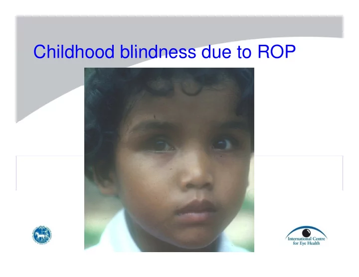
SLIDE 1
Childhood blindness due to ROP Childhood blindness due to ROP
SLIDE 2
Number of blind children/10 million pop, by cause and level of development by cause and level of development
SLIDE 3
Proportion of blindness due to ROP, by World Bank region by World Bank region
SLIDE 4
Estimates of numbers blind from ROP, b World Bank region Being re ised by World Bank region. Being revised…
Total >50 000 >50,000
SLIDE 5
Proportion of blindness due to ROP, b infant mortalit rates by infant mortality rates
SLIDE 6 ROP blindness – likely risk using IMR as a proxy 2010 proxy 2010
≤8/1000 Low risk of ROP blindness – good neonatal care and screening 9 – 60/1,000 High risk of ROP blindness – inadequate care and screening ≥61/1,000 Low risk of ROP – neonatal care not well developed
SLIDE 7
Retinal vascularisation during development
16 weeks GA 26 weeks GA 36 weeks GA 40 weeks GA
SLIDE 8 Pathogenesis of ROP
Relative hypoxia
SLIDE 9
Peripheral retinal hypoxia drives the ne blood essel gro th new blood vessel growth
SLIDE 10 Classification of ROP
u Site (zones and clock hours)
Severity (Stages)
u Severity (Stages) u Signs of BRB breakdown (“plus disease”)
Scarring
u Scarring
SLIDE 11 Classification of ROP - by zone (site) y ( )
Zone 3 Zone 2 Zone 1
SLIDE 12
Classification of ROP - by stage (severity)
Stage Features I Demarcation line II Ridge III Fibrovascular ridge IV Subtotal retinal detachment V Total retinal detachment
SLIDE 13
Stage 1 demarcation line Stage 1 demarcation line
SLIDE 14
Stage 1 demarcation line
SLIDE 15
Stage II ROP ridge Stage II ROP - ridge
SLIDE 16
Stage II ROP Stage II ROP
SLIDE 17
Stage III early Stage III - early
SLIDE 18
Stage III
SLIDE 19
Stage III ROP Stage III ROP
SLIDE 20
Stage III ROP g
SLIDE 21
Stage III ROP Stage III ROP
SLIDE 24 Stage 4 – subtotal retinal detachment
Courtesy Azad
SLIDE 25 Stage 4 – subtotal retinal detachment
Courtesy Azad
SLIDE 26
Stage V - total retinal detachment with open funnel with open funnel
SLIDE 27
Stage V - total retinal detachment ith l d f l with closed funnel
SLIDE 28
Stage V - inoperable retinal detachment g p
SLIDE 29
End stage eye blind from ROP g y
SLIDE 30
Child blind from ROP
SLIDE 31
Cicatricial disease with dragged vessels
SLIDE 32 Nat ral histor of ROP Natural history of ROP
u Disease starts 4-7 weeks after birth, and
progresses over the following few weeks
u Stage I and II disease
- spontaneous regression common
u Stage III “plus” disease (threshold disease)
- 50% progression to retinal detachment
u Stage IV and V disease
SLIDE 33 Classification of ROP
Classification of ROP - other
u “Plus” disease:
denotes breakdown of blood-ocular barriers, with pupil rigidity, dilated tortuous retinal vessels, vitreous haze
u Threshold disease:
5 + continuous clock hours of Stage III “plus” disease 8 h i t t l f St III “ l ” di
- r 8 hours in total of Stage III “plus” disease
SLIDE 34
“Plus” disease in posterior pole Plus disease in posterior pole
SLIDE 36
Changes to classification (2005) C a ges to c ass cat o ( 005)
u Pre-plus disease
Cl ifi ti f h t if di
u Clarification of how to assess if disease
is in zone 1
u Aggressive, posterior ROP (AP-ROP)
SLIDE 37
“Pre-plus” p disease
SLIDE 38 New stage: Aggressive posterior ROP (AP ROP) ROP (AP-ROP)
Courtesy Ells
SLIDE 39
New stage: Aggressive posterior ROP (AP-ROP) ROP (AP-ROP)
SLIDE 40
Indications for treatment - old Indications for treatment old
Threshold disease: A total of 8 discontinuous clock hours of stage III “plus” disease, or 5 or more continuous clock hours
SLIDE 41
Indications for treatment – new (earlier in the course of the disease) the course of the disease)
Type 1 pre-threshold disease:
– Zone 1, any ROP with plus disease (≥6 hours) – Zone 1, Stage 3 ROP +/- plus – Zone 2, Stages 2 or 3 with plus disease (≥6 h ) hours)
SLIDE 42 Rates of threshold disease ates o t es o d d sease
u Vary, depending on
– Case mix – Neonatal care and survival of most at risk – Screening criteria
u <1% in some UK units (<1,500g and/or <32
weeks)
u 15% in middle income countries (same criteria)
SLIDE 43
Treatment of threshold disease Treatment of threshold disease
Aim: confluent treatment of avascular retinal periphery with cryo or laser avoiding long ciliary vessels and with cryo or laser, avoiding long ciliary vessels and ridge
SLIDE 45
Indirect laser treatment
SLIDE 46
Baby receiving cryotherapy Baby receiving cryotherapy
SLIDE 47
Peripheral retinal cryo with laser Peripheral retinal cryo with laser
SLIDE 48
Disease regression after treatment Disease regression after treatment
SLIDE 49 Plus disease resolves with treatment
Before treatment At 2 weeks At 4 weeks
Courtesy Ells
SLIDE 50 Schematic representation of blindness d e to ROP in the West since 1940 due to ROP in the West since 1940
Oxygen restriction
Survival LBW babies
“first “second epidemic” epidemic”
Blindness due to ROP
1940 1950 1960 1970 1980 1990 2000 1940 1950 1960 1970 1980 1990 2000 BW: 1,000-1,500 gms 600-900 gms
SLIDE 51 Risk factors during the “first epidemic of ROP” in the West (1940s and 1950s) ROP” in the West (1940s and 1950s)
u Supplemental oxygen u No monitoring of blood gases u Birth weight: mean 1,300 (800 – 3,400 gs)
SLIDE 52 Risk factors during the “second epidemic
- f ROP” in the West (1970s on ards)
- f ROP” in the West (1970s onwards)
( 1 000
u Extremely low birth weight (<1,000 gms, av 750
gms) Extreme prematurity (<30 weeks GA: av 25
u Extreme prematurity (<30 weeks GA: av 25
weeks)
u Small for gestational age (SGA) u Small for gestational age (SGA) u Poor post natal weight gain u Fluctuating blood gases - hyperoxia/hypoxia
g g yp yp
u Factors predisposing to the “oxygen radical
disease of neonatology”
u [Ocular factors]
SLIDE 53 Characteristics of babies with “severe” ROP in UK USA and Canada ROP in UK, USA and Canada U K s c re e n in g
it i criteria Full term
Gilbert et al. Paediatrics 2005 115 518-525
SLIDE 54 Characteristics of babies with “severe” ROP in low/middle income countries
4000
Argentina (C ) Argentina (G) Argentina (L)
3000 3500
Argentina (L) Argentina (M) Argentina (P) Argentina (T) Brazil Chile Colombia Cuba Ecuador I di (D)
2000 2500 weight (gms)
India (D) India (H) India (M) Lithuania (K) Lithuania (V) Peru SS Peru Public Vietnam
1000 1500 Birth w 500 20 21 22 23 24 25 26 27 28 29 30 31 32 33 34 35 36 37 38 39 40
Gilbert et al. Paediatrics 2005 115 518-525
Gestational age (weeks)
SLIDE 55
Varying neonatal care in India - variation in exposure to risk factors for ROP
SLIDE 56 …variation in risk
Courtesy Ells
SLIDE 57 Risk factors during the “third epidemic
- f ROP” in middle income co ntries
- f ROP” in middle income countries
Historical perspective of ROP Historical perspective of ROP 1940-50s 1960-70s 1980s-present 1st epidemic 2nd epidemic Risk factors for ROP: Risk factors for ROP:
- prematurity
- low birth weight
- high oxygen
- illness factors
+ + ++++ + ++ ++ +++ + ++++ ++++ + +/- illness factors + + +/ <1,000 gms High mortality No ROP Mod mortality ROP + Low mortality ROP +++ I d V l 1,000-1,500 gms Improved Survival ROP +++ Low mortality ROP ++ Very low mortality No ROP Level of neonatal care Poor Moderate Excellent Level of neonatal care provided 3rd epidemic encompasses babies represented in all three columns
SLIDE 58 Risk factors during the “third epidemic
- f ROP” in middle income co ntries
- f ROP” in middle income countries
- Mixture of the first and second epidemic
- Different risk factors probably important in
p y p different clinical settings
- May be varying susceptibility in different
racial groups - blacks less susceptible
SLIDE 59 Summary of risk factors, and babies at risk of ROP babies at risk of ROP
- Varies, depending on neonatal outcomes:
– good neonatal outcomes: risk factors and babies g at risk similar to the West (i.e. extremely low birth weight; extreme prematurity; fluctuating oxygen levels etc) levels etc) – poor neonatal outcomes: risk factors similar to first epidemic (i.e. poorly controlled oxygen levels in more mature babies)
SLIDE 60 Prevention of blindness in children due to ROP due to ROP
– prevention of the disease from occurring in the first place first place
– early identification and treatment to prevent the – early identification and treatment, to prevent the consequences of the disease
y p
– Interventions to restore function
SLIDE 61 Primary prevention of ROP - 1 Primary prevention of ROP - 1
– avoid unnecessary Caesarian sections d t t l – good antenatal care – prevent teenage pregnancies (26% mothers <20 years old in a recent screening prog study <20 years old in a recent screening prog study in Ecuador) – prevent multiple birth (e.g. from IVF) prevent multiple birth (e.g. from IVF) – good obstetric care
SLIDE 62 Primary prevention of ROP - 2 Excellent neonatal care
Excellent neonatal care
– monitoring blood gases – systemic steroids prior to preterm delivery systemic steroids prior to preterm delivery
– surfactants surfactants – vitamin E
Ineffective: – Light restriction – Vitamin A supplementation Vitamin A supplementation
SLIDE 63 Secondary prevention: Secondary prevention:
– screening to identify babies with threshold, or pre threshold disease pre threshold disease – treatment by peripheral retinal ablation by cryotherapy or laser cryotherapy or laser – increasing oxygen saturation in babies with threshold disease gave essentially negative findings (STOP-ROP trial)
SLIDE 64 Screening for threshold ROP g
- Is a screening programme needed?
– Only if there is intensive neonatal care – Only if there is intensive neonatal care
- Which units should be included?
– Start in larger units where at risk babies are surviving Start in larger units where at risk babies are surviving
– ? <2,000 gms and/or <32 weeks + “sickness” ? 2,000 gms and/or 32 weeks sickness
– First examination 4 weeks after birth
– Indirect ophthalmoscopy with dilated pupil p py p p – +/- lid speculum, with depressor to rotate the eye
SLIDE 65 Indirect ophthalmoscopy in the neonatal unit neonatal unit
Courtesy Ells
Courtesy Zin
SLIDE 66 Screening for ROP g
u Who?
– Skilled ophthalmologist (VR, or paediatric)
u How often:
Every 1 or 2 weeks depending on degree of prematurity and – Every 1 or 2 weeks, depending on degree of prematurity, and findings at each examination
u For how long?
U til i / l i ti – Until regression / vascularisation
u How should this be organised?
– Neonatologist is responsible for identifying babies to be examined – Diary system useful – Nurse dilates the pupils – Regular visits, on a pre-determined day and time (for discharged babies to be brought back)
SLIDE 67 New developments in screening: digital imaging digital imaging
Courtesy Ells
SLIDE 68 RetCam RetCam
- RetCam can be used
- 1. As an adjunct to indirect ophthalmoscope
– advantage: can record image
- 2. For telemedicine screening with a) a technician
who takes and grades images there and then, or b) uploaded images onto the internet Images have to uploaded images onto the internet. Images have to be interpreted by remote experts within 48 hours. For each baby have to decide: a) discharge b) y ) g ) follow up (& when) c) needs treatment
- Telemedicine screening: still experimental
SLIDE 69 New concepts in screening: WINROP
- Weight gain during first few weeks of life
predicts subsequent ROP risk
- Mediated via IGF-1
- WINROP: a computer model developed in
S d It d t b lid t d i tti
- Sweden. It needs to be validated in settings
where bigger babies are also developing severe ROP severe ROP
SLIDE 70 Treatment of ROP Treatment of ROP
u Indications: u Indications:
– Prethreshold disease in one or both eyes
u How:
– Laser (or cryotherapy if laser not available)
u Aim of treatment:
– Complete ablation of avascular retinal periphery
u Anaesthesia:
– Sedation + analgesia or GA
u Post op:
– Mydriatics and topical steroids
SLIDE 71 Follow up Follow up
- All babies who have been treated, to ensure
regression
– If not regressed, retreat
- Premature babies with or without ROP have a
markedly increased risk of the following:
i ifi t f ti – significant refractive errors – strabismus cortical visual impairment – cortical visual impairment – disorders of higher visual pathways – optic atrophy and hypoplasia
- ptic atrophy and hypoplasia
SLIDE 72 Tertiary prevention of ROP Tertiary prevention of ROP
- Vitreoretinal surgery for Stage IV and Stage V:
– no randomised clinical trials have been done – some surgeons believe in retinal detachment surgery for Stage IV most surgeons do not now operate on Stage V – most surgeons do not now operate on Stage V, as the surgery is so difficult, and the functional results are poor
- Rehabilitation, special education, support
services
SLIDE 73 Potential new treatment for ROP
- Anti-VEGF preparations by intravitreal injection
Anti VEGF preparations by intravitreal injection
- Effective as a “salvage” treatment
- Some advocate it as first line treatment
Some advocate it as first line treatment
– is absorbed systemically y y – not known what effect it might have on developing vasculature elsewhere, glomeruli and alveoli
- Clinical trial on-going in US BUT not investigating
long term complications
SLIDE 74 Programmes for ROP Programmes for ROP
Need good coverage
- Need good management information systems
e.g. online data recording for each baby
- Need to be co-ordinated and monitored
- Need financial support, ideally from Ministries of
Health Health
- Need to involve parents
- May need
ay eed – training – equipment