
A transcriptional sketch of a human breast A transcriptional sketch of a human breast cancer by 454 deep sequencing cancer by 454 deep sequencing
The cancer transcriptome is difficult to explore, due to the heterogeneity of quantitative and qualitative transcriptional events linked to the disease status. An increasing number of “unconventional” transcripts, such as novel isoforms, noncoding RNAs, somatic gene fusions and deletions have been associated with the tumoral state. Massively parallel sequencing techniques make full- transcriptome sequencing feasible with a limited laboratory and financial effort, and provide a framework for exploring the complexity inherent to the cancer transcriptome.
We developed a 454 deep sequencing and bioinformatics analysis protocol to investigate the molecular composition of a breast cancer poly(A)+ transcriptome. This method utilizes a normalization step to diminish the abundance of highly expressed transcripts and biology-oriented bioinformatic analyses to facilitate detection of rare and novel transcripts, which may enhance our understanding of the aetiology of the disease. We demonstrate that combining 454 deep sequencing with a normalization step and careful bioinformatic analysis facilitates the discovery of rare transcripts, and can be used as a qualitative tool to characterize transcriptome complexity, revealing many hitherto unknown transcripts, splice isoforms, and gene fusion events, even at a relatively low sequence sampling.
Library normalization: wet lab matters Library normalization: wet lab matters Reference Gene1 454 Reads mapped UniGene ESTs Probability of to the genome differential (39,700) (194,806) expression between the libraries ACTB 11 187 Prob > 0.999 GAPDH 31 225 Prob > 0.999 HPRT1 7 0 0.5 < Prob < 0.6
The sequence landscape The sequence landscape (left) Distribution of sequence lengths shows a good approximation to a Normal Distribution (right) Sequence reads show an higher representation toward the 3’end of a transcript, but coverage is present along all the transcript
HPC challenges (and solutions) HPC challenges (and solutions) (1) Data distribution: split the dataset on chunks of, e.g., 2.000 sequences each. (2) Memory management may be very problematic with short word search parameters (- W 4 for Blast or – tileSize 8 for Blat) when comparing against the large and repetitive human or mouse genomes. (3) String-based searches eat up lots of disk space and processor time quickly => we used two bioinformatic clusters and an eight-processor server with large shared memory (8 Giga) www.litbio.org www.vital-it.ch
Mapping to the Genome and the Mapping to the Genome and the Transcriptome Transcriptome Set Description Number of reads Total (unfiltered) 251.262 Mapping to the genome, 70% 194.806 coverage, high stringency Subset with a single match on the 132.113 reference dataset genome at 98% identity and 98% coverage (98.98.1 dataset) Subset with a single match on the 114.427 – 87 % of the reference genome and 100% coverage of the dataset alignment Subset of 98.98.1 dataset matching 59.632 - – 45 % of the reference with max 6 errors (mismacthes + dataset indels) and 90% coverage on UCSC all_mrna and RefSeq – canonical transcripts dataset Subset of 98.98.1 dataset matching 118.840 - – 90 % of the reference inside an UCSC Known Gene dataset (Intragenic dataset, intronic + exonic transcripts) Matching with max 6 errors 68.396 – 52 % of the reference (mismacthes + indels) and 90% dataset coverage to the Human ORESTES EST dataset (764.587 sequences)
Classification of the sequences in Classification of the sequences in the genome annotation context the genome annotation context Sequence class Number of reads Intergenic Unspliced 6.298 Intergenic Spliced 402 Intragenic Unspliced – total 97.690 3 TERM 2.475 (Poli-A) (989) (INTERNAL) (1.486) 5 TERM 2.807 (TSS) (1.113) (INTERNAL) (1.694) EXON 1.331 INTRAEXON 64.326 INTRON 26.751 Intragenic Spliced 10.037 Total 114.427 Abbreviations: 3 TERM, read which extend the annotated 3 ’ term of the target gene. Poli-A: read which extends at 3 ’ the last exon. INTERNAL: read which extends at 3 ’ any exon except the last. 5 TERM: read which extend the annotated 5 ’ term of the target gene. TSS: read which extends at 5 ’ the first exon. INTERNAL: read which extends at 5 ’ any exon except the first. EXON: read mapping inside an exon with one or both ends coincident with exon boundaries. INTRAEXON: read mapping completely inside an exon of the target gene. INTRON: read mapping completely inside an intron.
Genes or Transcripts Fusions Genes or Transcripts Fusions in solid tumors in solid tumors Nature Reviews Cancer 7, 233-245 (April 2007) The impact of translocations and gene fusions on cancer causation Felix Mitelman Bertil Johansson & Fredrik Merten Abstract Chromosome aberrations, in particular translocations and their corresponding gene fusions, have an important role in the initial steps of tumorigenesis; at present, 358 gene fusions involving 337 different genes have been identified. An increasing number of gene fusions are being recognized as important diagnostic and prognostic parameters in malignant haematological disorders and childhood sarcomas. The biological and clinical impact of gene fusions in the more common solid tumour types has been less appreciated. However, an analysis of available data shows that gene fusions occur in all malignancies, and that they account for 20% of human cancer morbidity. With the advent of new and powerful investigative tools that enable the detection of cytogenetically cryptic rearrangements, this proportion is likely to increase substantially.
Genes or Transcripts Fusions Genes or Transcripts Fusions Perfect Fusion of two sequences located in different chromosomes
Genes or Transcripts Fusions Genes or Transcripts Fusions UBR4 , commonly known as p600 or retinoblastoma protein-associated factor 600, is a cellular target of the human papillomavirus type 16 E7 oncoprotein that regulates cellular pathways, contributing to anchorage-independent growth and cellular transformation. UBR4-E7 interaction strongly contributes to cellular transformation (Huh 2005). The GLB1 gene encodes beta-galactosidase-1 (EC 3.2.1.23), a lysosomal hydrolase that cleaves the terminal beta-galactose from ganglioside substrates and other glycoconjugates. The predicted fusion, verified by direct sequencing of the original cDNA library, links exon 16 of the gene UBR4 with the terminal exon (coding + 3’UTR), common to all the transcript variants, of the GLB1 gene. Our sequence ( 4A ) is colinear with both transcripts and exon-exon junctions are clear in the hybrid sequence. The predicted final processed fusion cDNA UBR4/GLB1 would be 14,022-bp long and would produce a very large protein of 4.526 residues, which however is shorter than the original UBR4 protein (5.183 residues).
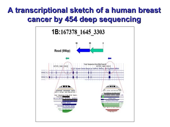
A Genomic Deletion ? A Genomic Deletion ? An example of a transcriptional or genomic deletion event is provided by the sequence read 1B (167378_1645_3303), located on chromosome 8. We interpret this sequence as a deletion, probably due to a loop which causes the inclusion of exons 2 and 7 of the WHSC1L1 gene in an inverted order in the mature transcript. This transcript was also confirmed by direct sequencing of the cDNA library. The WHSC1L1 gene is related to the Wolf-Hirschhorn syndrome candidate- 1 gene and encodes a protein with PWWP (proline-tryptophan-tryptophan- proline) domains. Two alternatively spliced WHSC1L1 variants have been described. The long isoform contains a PHD-finger domain (an interleaved type of Zn finger chelating 2 Zn ions) and a SET domain (protein-protein interaction domain); however, the function of the protein has not been determined yet and hence the relevance to cancer aetiology of this deletion is uncertain.
Rare and Novel Isoforms Rare and Novel Isoforms The following examples clearly demonstrate that it is possible to identify known, novel and even possible cancer-related isoforms with this sequence length and at this sequencing depth. We were able to retrieve known isoforms of IGL@ (Unigene cluster 449585 Immunoglobulin lambda joining 3), which is located in an area of very active and complex genomic rearrangements. The corresponding read 045624_1590_1179 (which we renamed 6B ) is a 102 nt transcript fragment which maps to the Variable, Light and Join segments of immunoglobulin genes on Chr 22q11.1-q11.2. When mapped to the genome this read aligns to the first 60 nucleotides, with 98% identity, to nt 21571798-21573177 (divided in two exons) and with nt 60-101, with 95% identity, to nt 21060830-21060871 (third exon) of the minus strand of Chr 22. Hence this single 102 nt read contains three different exons, the second and the third separated by around 511.000 bases.
Recommend
More recommend