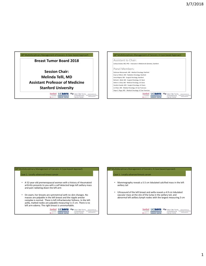

3/7/2018 18 th Multidisciplinary Management of Cancers: A Case‐based Approach 18 th Multidisciplinary Management of Cancers: A Case‐based Approach Breast Tumor Board 2018 Assistant to Chair: Joshua Gruber, MD, PhD – Instructor in Medicine & Genetics, Stanford Panel Members: Session Chair: Suleiman Massarweh, MD ‐ Medical Oncology, Stanford Erqi Liu Pollom, MD ‐ Radiation Oncology, Stanford Melinda Telli, MD Irene Wapnir, MD ‐ Surgical Oncology, Stanford Richard J. Bold, MD ‐ Surgical Oncology, UC Davis Assistant Professor of Medicine Helen K. Chew, MD ‐ Medical Oncology, UC Davis Candice Sauder, MD ‐ Surgical Oncology, UC Davis Stanford University Jo Chien, MD ‐ Medical Oncology, UC San Francisco Hope S. Rugo, MD – Medical Oncology, UC San Francisco 18 th Multidisciplinary Management of Cancers: A Case‐based Approach 18 th Multidisciplinary Management of Cancers: A Case‐based Approach Case 1: Locally advanced breast cancer Case 1: Locally advanced breast cancer • • A 52‐year‐old premenopausal woman with a history of rheumatoid Mammography reveals a 5.5 cm lobulated calcified mass in the left arthritis presents to you with a self detected large left axillary mass axillary tail and pain radiating down the left arm • Ultrasound of the left breast and axilla reveals a 4.9 cm lobulated • On exam, her breasts are symmetrical with no skin changes. No vascular mass at the site of the lump in the axillary tail, and masses are palpable in the left breast and the nipple areolar abnormal left axillary lymph nodes with the largest measuring 3 cm complex is normal. There is left infraclavicular fullness. In the left axilla, matted nodes are palpable measuring 5 x 5 cm. There is no left arm edema. The right breast is unremarkable. 1
3/7/2018 18 th Multidisciplinary Management of Cancers: A Case‐based Approach 18 th Multidisciplinary Management of Cancers: A Case‐based Approach Case 1: Mammography Case 1: Axillary ultrasound 18 th Multidisciplinary Management of Cancers: A Case‐based Approach 18 th Multidisciplinary Management of Cancers: A Case‐based Approach Case 1: Locally advanced breast cancer Case 1: Locally advanced HER2‐positive breast cancer • A breast MRI is ordered and reveals multiple left axillary masses, • A core biopsy of axillary mass is pursued and reveals: infraclavicular and supraclavicular nodes. No abnormal enhancement is seen in left breast. Right breast is benign . • Poorly differentiated metastatic carcinoma most c/w breast origin in an axillary lymph node • It is favored that the 4.7 cm dominant axillary mass at 2:30, 15cm from the • ER 2%, PR 2%, HER2 3+ via IHC nipple, represents the primary breast carcinoma. • Ki‐67 = 40% • A staging PET/CT scan reveals: • She is staged as having TX N3c M0 Clinical Stage IIIC disease • Bulky FDG avid nodes in left axilla, subpectoral, and supraclavicular areas • Primary breast cancer not visualized • No distant metastases 2
3/7/2018 18 th Multidisciplinary Management of Cancers: A Case‐based Approach O 18 th Multidisciplinary Management of Cancers: A Case‐based Approach E Case 1: Anatomic N1 N2a N2b Case 1: Treatment of locally advanced HER2‐positive breast cancer Fixed/matted S nodal stage is N3c nodal mass U • She is treated with neoadjuvant docetaxel, carboplatin, trastuzumab and pertuzumab L (TCH+P) for 6 cycles A N • Palpable residual disease remains after chemotherapy clinically. O S N3a N3b N3c • A repeat breast MRI reveals: R • The largest axillary lesion decreased from 49 to 23 mm E P • All lesions decreased in size consistent with treatment response R • No new lesions O • No left breast primary tumor identified AJCC Staging Manual 8 th edition, 2017 F Breast chapter Hortobagyi et al. 18 th Multidisciplinary Management of Cancers: A Case‐based Approach 18 th Multidisciplinary Management of Cancers: A Case‐based Approach Case 1: For surgical management, you recommend: Case 1: Surgery • She undergoes bilateral breast reduction with left axillary nodal 1. Mastectomy and ALND dissection 2. ALND only • Pathology reveals: • No evidence of carcinoma or treatment effect in the breast • 25/33 nodes involved with residual carcinoma 3. Lumpectomy and ALND • Extensive extra‐capsular extension • ER‐negative, PR‐negative • HER2‐positive (IHC 2+, FISH ratio 1.42, HER2 copies/cell = 7.4, AMPLIFIED) • Ki‐67 40% 3
3/7/2018 18 th Multidisciplinary Management of Cancers: A Case‐based Approach 18 th Multidisciplinary Management of Cancers: A Case‐based Approach Case 1: Regarding adjuvant radiotherapy, you recommend: Case 1: Regarding adjuvant systemic therapy, you recommend: 1. Adjuvant trastuzumab 1. Whole breast and regional nodal irradiation 2. Adjuvant trastuzumab + pertuzumab 2. Regional nodal irradiation only 3. Adjuvant capecitabine + trastuzumab 4. Adjuvant capecitabine + trastuzumab + pertuzumab 5. Adjuvant trastuzumab followed by adjuvant neratinib for one year 18 th Multidisciplinary Management of Cancers: A Case‐based Approach 18 th Multidisciplinary Management of Cancers: A Case‐based Approach ExteNET – Study Design Case 1: APHINITY adjuvant HER2+ (trastuzumab + pertuzumab/placebo for 1 year) Case 1: Early Stage Breast Cancer Primary Exploratory Analysis Analysis HER2+ 1:1 RANDOMIZATION Neratinib x 1 yr 2-year follow-up for iDFS 5-year follow-up for iDFS 240 mg/day Overall survival Stratification Factors: N=1420 • Nodes 0, 1-3, vs 4+ • ER/PR status Placebo x 1 yr • Concurrent vs sequential N=1420 trastuzumab N=2840 von Minckwitz et al. NEJM 2017 4
3/7/2018 18 th Multidisciplinary Management of Cancers: A Case‐based Approach 18 th Multidisciplinary Management of Cancers: A Case‐based Approach Case 1: ExteNET primary analysis Case 1: Adjuvant treatment • She completes whole breast and regional nodal irradiation with concurrent capecitabine Neratinib Placebo (N=1420) (N=1420) • She completes 6 months of adjuvant capecitabine and one year of iDFS Events 67 (4.7%) 106 (7.5%) trastuzumab + pertuzumab 2-year KM estimate 94.2% 91.9% Difference (95% CI) 2.3% (0.3%, 4.3%) Stratified log-rank p-value 0.008 (two-sided) • She declines endocrine therapy and neratinib Stratified HR (95% CI) 0.66 (0.49, 0.90) • She remains NED 18 th Multidisciplinary Management of Cancers: A Case‐based Approach 18 th Multidisciplinary Management of Cancers: A Case‐based Approach Case 2: Male Breast Cancer Case 1: • A 72‐year‐old man presents with a lump in the right breast and skin changes involving the nipple and areola. • Family history is significant for ovarian cancer in his mother at age 80 and early onset breast cancer in a maternal first cousin at age 30. END OF CASE 1 • On exam, he has a 4 x 5 cm right retroareolar mass with nipple retraction and matted right axillary nodes measuring 4 cm. Dermal involvement by carcinoma is noted. • An ultrasound is ordered and reveals: • 3.9 cm retroareolar mass at 2 o’clock • Multiple right axillary nodes up to 2.9 cm 5
3/7/2018 18 th Multidisciplinary Management of Cancers: A Case‐based Approach 18 th Multidisciplinary Management of Cancers: A Case‐based Approach Case 2: Male Breast Cancer Case 2: Male Breast Cancer ‐‐ RISK FACTORS • Right breast core biopsy is pursued and reveals: • 0.7% of all breast cancer diagnoses • IDC grade 3 • Testicular dysfunction (crytporchidism, orchitis, infertility) • ER 99% • PR 99% • Klinefelter’s syndrome (XXY): 3‐7% (50‐fold increase) • HER2 negative (DISH ratio 1.2) • Family history of female breast cancer (2.5‐fold increase risk) • Right axillary node core biopsy reveals metastatic carcinoma • Prior radiation therapy to the chest • Genetic predisposition • CT CAP and bone scan negative for distant metastases • Clinical stage: cT4 N2 M0 ‐‐ Stage IIIB Giordano SH The Oncologist 2005 18 th Multidisciplinary Management of Cancers: A Case‐based Approach 18 th Multidisciplinary Management of Cancers: A Case‐based Approach Case 2: Male Breast Cancer Case 2: Male Breast Cancer • Mammogram post‐neoadjuvant • He declines genetic testing chemotherapy reveals interval decrease in the size of breast mass and axillary • Neoadjuvant chemotherapy is pursued with doxorubicin and disease cyclophosphamide (AC) x 4 followed by paclitaxel weekly x 12 • Mass 3.6 ‐> 3.2 cm • Largest axillary node 2.8 ‐> 1.7 cm • Clinically, the NAC is retracted with no dominant mass and he no longer has palpable axillary adenopathy 6
Recommend
More recommend