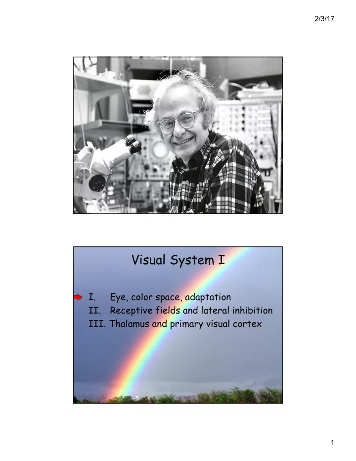

2/3/17 1 Visual System I I. Eye, color space, adaptation II. Receptive fields and lateral inhibition III. Thalamus and primary visual cortex 2 1
2/3/17 Window of the Soul 3 Information Flow: From Photoreceptors to the Optic Nerve Layer 1: Photoreceptors Layer 2: Bipolar cells Layer 3: Ganglion cells Stimulus: Light Output: Optic Nerve 2
2/3/17 Types of Photoreceptors Rods are specialized to Cones are specialized detect in dim light, for detecting color and do not detect color and bright light Disks containing photopigment (light absorber) 5 The Distribution of Rods and Cones Differs Across the Retina fovea 20 X more rods than cones Rods Cones -Periphery -Fovea -Dim light -Bright light -Black & white -Color -Signal amplification -Little pooling -Much pooling (fine discrimination ) (noise reduction) 6 3
2/3/17 7 Red is important! 8 4
2/3/17 Cone tuning S curves M L 9 Color perception is based on the relative activation of the three cone types—a single cone type carries NO color information 10 5
2/3/17 Cone color space 11 Adaptation: decreasing response to a constant stimulus Firing rate code Edgar Adrian “The Basis of Sensation” 1928 6
2/3/17 13 14 7
2/3/17 15 16 8
2/3/17 17 Visual System I I. Eye, color space, adaptation II. Receptive fields and lateral inhibition III. Thalamus and primary visual cortex 18 9
2/3/17 9 Amount of light from the white of a 10 newspaper viewed in direct sunlight 8 10 Amount of light from the black text of a newspaper viewed in direct sunlight 7 10 6 (relative units) 10 Luminance 5 10 4 10 3 10 Amount of light from the white of a 2 10 newspaper viewed indoors 10 1 Amount of light from the black text of a newspaper viewed indoors 19 20 10
2/3/17 21 Receptive Field: An area in which stimulation leads to response of a particular sensory neuron. 22 11
2/3/17 Antagonistic center-surround spatial receptive fields 2 classes of cells: On-center: Off-center: prefer light at center prefer dark at center and and dark in surround light in surround Receptive Field of Retinal Bipolar Neurons = the area of the retina that, when stimulated with light, influences the activity of a bipolar cell Center Surround: these cells send lateral inhibition to our bipolar cell (indirect) Surround portion of RF Center portion of RF (direct) Bipolar cell 24 12
2/3/17 Retinal ganglion cells also fire according to a center-surround organization 25 Mach Bands 13
2/3/17 Lateral inhibition produces contrast enhancement “...at the expense of fidelity of representation.” Lateral inhibition produces center- surround RFs, which produce contrast enhancement 28 14
2/3/17 Eye Summary • Sensory responses depend on the recent history of stimulation, due to adaptation effects • Bipolar cells integrate a neighborhood of photoreceptor responses to exhibit antagonistic center-surround receptive fields • Retinal ganglion cells integrate bipolar responses and send action potentials into brain • Center-surround RFs (lateral inhibition) enhance spatial contrasts • Luminance and color information are processed in parallel by different cells 29 Visual System I I. Eye, color space, adaptation II. Receptive fields and lateral inhibition III. Thalamus and primary visual cortex 30 15
2/3/17 Eye to Brain 32 16
2/3/17 Left field Right field Figure 10.3 33 Retinal Inputs to the Right and Left LGN 34 17
2/3/17 “Geniculate” means bent like a knee 35 Figure 10.4b 36 18
2/3/17 The striate cortex (V1) contains a retinotopic map 37 Spatial Receptive Fields Spatial receptive field = region of visual field to which a cell responds Receptive field of this V1 cell is to the left of where the eyes are looking (Green = locations where light would cause cell to be excited Red = locations where light would cause cell to be inhibited) Cell in V1 38 19
2/3/17 Shapes of Spatial Receptive Fields From the Eye to Primary Visual Cortex Flow of information: Photoreceptors + Bipolar cells + - - - Ganglion Cells + - - - - - - + LGN Cells - - - - - + - - V1 Cells 39 Tuning Curves in the Primary Visual Cortex Single neuron: average firing rate (Hz) orientation (deg) Many neurons: 60 rate (Hz) 0 -180 0 180 orientation angle in degrees 20
2/3/17 Simple Feedforward Network: How V1 Receptive Fields are Formed from LGN RF’s LGN Receptive cells fields of LGN cells V1 cell that responds best to a bar oriented at 45 0 Simple Feedforward Network: How V1 Receptive Fields are Formed from LGN RF’s LGN Receptive cells fields of LGN cells V1 cell that responds best to a bar oriented at 45 0 Hubel & Wiesel, 1962 21
2/3/17 Conclusions • Receptive fields and tuning curves characterize the response properties of sensory neurons • Visual neurons are arranged in retinotopic “maps” • Different sensory features are processed in parallel in different brain areas • More complex and specific features- sensitivities are constructed from lower- level features (hierarchy) 43 22
Recommend
More recommend