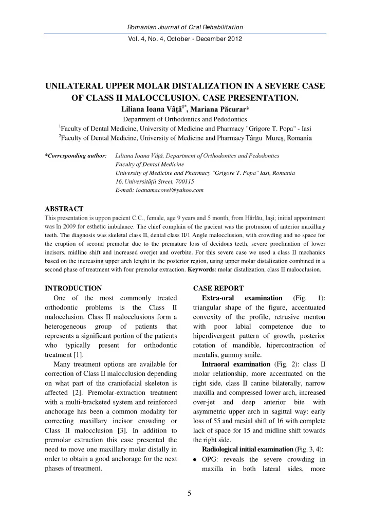

Romanian Journal of Oral Rehabilitation Vol. 4, No. 4, October - December 2012 UNILATERAL UPPER MOLAR DISTALIZATION IN A SEVERE CASE OF CLASS II MALOCCLUSION. CASE PRESENTATION. 1* Department of Orthodontics and Pedodontics 1 Faculty of Dental Medicine, University of Medicine and Pharmacy "Grigore T. Popa" - Iasi 2 Faculty of Dental Medicine, University of Medicine and Pharmacy *Corresponding author: Faculty of Dental Medicine University of Medicine and Pharmacy "Grigore T. Popa" Iasi, Romania 16, ii Street, 700115 E-mail: ioanamacovei@yahoo.com ABSTRACT imbalance. The chief complain of the pacient was the protrusion of anterior maxillary teeth. The diagnosis was skeletal class II, dental class II/1 Angle malocclusion, with crowding and no space for the eruption of second premolar due to the premature loss of decidous teeth, severe proclination of lower incisors, midline shift and increased overjet and overbite. For this severe case we used a class II mechanics based on the increasing upper arch lenght in the posterior region, using upper molar distalization combined in a second phase of treatment with four premolar extraction. Keywords : molar distalization, class II malocclusion. INTRODUCTION CASE REPORT One of the most commonly treated Extra-oral examination (Fig. 1): orthodontic problems is the Class II triangular shape of the figure, accentuated malocclusion. Class II malocclusions form a convexity of the profile, retrusive menton heterogeneous group of patients that with poor labial competence due to represents a significant portion of the patients hiperdivergent pattern of growth, posterior who typically present for orthodontic rotation of mandible, hipercontraction of treatment [1]. mentalis, gummy smile. Many treatment options are available for Intraoral examination (Fig. 2): class II correction of Class II malocclusion depending molar relationship, more accentuated on the on what part of the craniofacial skeleton is right side, class II canine bilaterally, narrow affected [2]. Premolar-extraction treatment maxilla and compressed lower arch, increased with a multi-bracketed system and reinforced over-jet and deep anterior bite with anchorage has been a common modality for asymmetric upper arch in sagittal way: early correcting maxillary incisor crowding or loss of 55 and mesial shift of 16 with complete Class II malocclusion [3]. In addition to lack of space for 15 and midline shift towards premolar extraction this case presented the the right side. need to move one maxillary molar distally in Radiological initial examination (Fig. 3, 4): order to obtain a good anchorage for the next OPG: reveals the severe crowding in phases of treatment. maxilla in both lateral sides, more 5
Romanian Journal of Oral Rehabilitation Vol. 4, No. 4, October - December 2012 accentuated on the right quadrant, with an Diagnosis: increased mesial tip of 16 and contact Dental: class II/1 Angle maloclusion, with between 16 and 14; tendency of 13, 23 to crowding located in both arches more be impacted; in mandible it is evident that severe in the right upper lateral quadrant lower incisors are very proclined and also with no space for the eruption of 15 due to there is a lack of space for premolars, the premature loss of deciduous teeth, especially on the left side. reduced apical base in maxilla and severe Profile cephalometry: reveals the severe proclination of lover incisors, midline shift hiperdivergence (Sn/GoGn = = and increased over-jet and overbite. Skeletal: class II, retrognatic mandible, and the proclination of lower incisors hiperdivergent pattern. Fig. 1. Extraoral examination Fig. 2. Intraoral examination Fig. 3. Initial OPG 6
Romanian Journal of Oral Rehabilitation Vol. 4, No. 4, October - December 2012 Fig. 4. Cephalometric analysis Treatment plan: applied a transpalatal bar for anchorage Initial objectives: space regaining in the Overall treatment plan: extractional case upper arch in order to ensure an critical (14, 24, 34, 44) with critical anchorage in anchorage during incisor retraction maxilla and maximal anchorage in Initial mandible, using Roth slot 0.22 preadjusted treatment: upper first molar fixed appliance. Total duration of distalization on the right side treatment: 3 years (2009-2012). Appliance: palatal plate with distalization screw RESULTS Type of distalization: intramaxillary Upper molar distalization and space appliance with palatal point of force opened are illustrated in fig. 5 and 6. application. Time for distalization: 7 month. After molar distalization we Fig. 5. Palatal plate and the space between 14 and 16 Fig. 6. Fixed appliance in lower arch and transpalatal bar in upper arch after 16 distalization 7
Romanian Journal of Oral Rehabilitation Vol. 4, No. 4, October - December 2012 Comparative OPG analysis at T1 and T2 first molar is distalized on lateral cephalogram improved tip forward for this tooth (Fig. 7). mm, upper incisors are proclined during At the end of the overall treatment it is distalization and retroclined after premolar evident that a good facial aesthetics is extraction, and the occlusal angle decreased obtained (Fig. 8, 9, 10) and cephalometric values, especially for lower incisors, overjet after the extraction of the four premolars. and overbite are corrected (Table 1). Upper T1 T2 Variables predistalization postdistalization M1 S Md dr 113 110 M1 S Md stg 104 104 Ang M1 S dr 12 9 Ang M1 S stg 3 3 R1S dr 15/15 15/7 R1S stg 13/13 14/9 Fig. 7. Comparative OPG analysis for upper molar distalization Fig. 8. Profile changes: final (2012) Fig. 9. Cephalometric evaluation at the end of treatment vs. initial (2009) (2012) Fig. 10. Extraoral and intraoral aspects at the end of the treatment 8
Romanian Journal of Oral Rehabilitation Vol. 4, No. 4, October - December 2012 I. Aesthetics Variables Pre-treatment Intermediary (post distalization) Post-treatment (post extractional) Lsup-E -2 -3 -6 Linf-E 0 -1 -3 18 16 14 TC UL 10 12 14 Angle Z 67 68 68 Nazio-labial angle 112 113 105 II. Skeletal Variables Pre-treatment Intermediary (post distalization) Post-treatment (post extractional) Sagittal SNA 83 82 80 SNB 75 74 72 ANB 8 8 8 A-PTV 55 55 53 B-PTV 47 48 48 Vertical SN/GoGn 38 38 38 FMA 33 33 33 11 11 13 SN/PP SN/Pocl 24 20 27 HFA 67 66 66 HFP 40 42 42 Ip/a 0.59 0.63 0.63 III. Dental Pre-treatment Intermediary (post distalization) Post-treatment (post extractional) Variables Angular Ax Isup-SN 92 94 90 Ax Pm1-SN 70 78.5 - 72 67 70 Ax M1-SN Ax M2-SN 80 74 65 Ax M3-SN 65 61 53 Ax Isup-NA 10 12 10 Ax Iinf-NB 35 28 30 IMPA 102 97 97 FMIA 45 50 50 AxM1inf-GoGn 92 90 102 Ii 128 132 132 Linear horizontal Isup-PTV 60 61 55 U1-NA 3 4 0 Pm1-PTV 31 32,5 - U6-PTV 16 14,5 20 M2-PTV 9 8 12 M3-PTV 0 0 5 Iinf-PTV 56.5 54.5 53 L1-NB 10 8 7 L6-PTV 15 16 19 OJ 3,5 6.5 2 Linear vertical Isup-PP 29 28 27 Pm1-PP 23 24 - 21 22.5 20 U6-PP M2-PP 12 16 16 M1inf-GoGn 30 30 28 Iinf-GoGn 38 40 34 OB 3 5 1 Table 1. Comparative changes pre-treatment, postdistalization and at the end of extractional treatment 9
Romanian Journal of Oral Rehabilitation Vol. 4, No. 4, October - December 2012 15-16 activated from class II elastics; elastic DISCUSSIONS For extreme cases we used a class II chain for upper canines distalization. And in mechanics based on the increasing upper arch the mandible: anchorage preparation: molar tip-back; Stabilization 0.19 x 0.25 stainless length in the posterior region, using molar steel lower archwire [6]. distalization [4]. Aesthetic criteria determines in severe class II malocclusions the extraction of four premolars, lingual displacement of the CONCLUSIONS lower anterior teeth and retraction of the When crowding in the maxilla is entire upper arch related to the mandibular associated with Class II molar and skeletal arch [5]. relationship maxillary molar distalization can In this case we used the lower arch as be performed to increase the anchorage. Then, anchorage unit during upper incisors the molars are held in place whereas canines retraction and the upper arch with critical and incisors usually are retracted by anchorage to minimize the side effects of this conventional multi-bracket techniques. Molar retraction. distalization, especially in the maxilla, is often The technical orthodontic measures a challenge in orthodontic treatment . In this involves in the maxilla the use of: distalizing case we presented a combination therapy, upper 0.18 x 0.25 stainless steel arch with where maxillary molar distalization is omega loop placed in front of 16, 26; palatal associated in cases with crowding with dental plate and transpalatal bar for upper molar extraction in a severe class II molar and distalization; class II elastics for maxillary skeletal relationship. retraction; compression coil spring between REFERENCES 1. Karlsson I, Bondemark L. Intraoral maxillary molar distalization. Angle Orthod 2006;76:923-9. 2. Proffit W., H.W. Fields. Contemporary Orthodontics. Third Ed. Mosby 2000, 478-508 3. Herb Klontz. The Extraction / nonextraction dilemma the Class II solution The Tweed Profile 2006;5:25-30 4. Horn A. Advanced Course of Edgewise technic; Bucharest, 2003 5. Merrifield L. The profile lines as an aid in critically evaluating facial esthetics. Am. J. Orthod. Dento- Facial Orthop; 1966;52;11;804-822 6. Marcotte. M. Biomechanics in Orthodontics. B. C. Decker, Inc., Toronto, Philadelphia, 1990, 127- 137 10
Recommend
More recommend