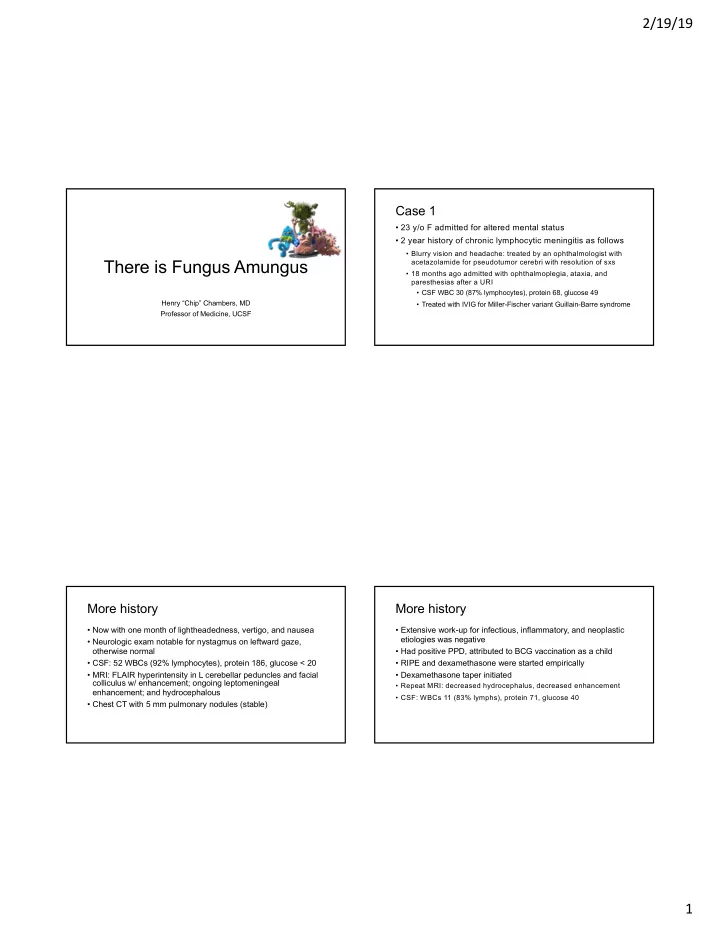

2/19/19 Case 1 • 23 y/o F admitted for altered mental status • 2 year history of chronic lymphocytic meningitis as follows • Blurry vision and headache: treated by an ophthalmologist with There is Fungus Amungus acetazolamide for pseudotumor cerebri with resolution of sxs • 18 months ago admitted with ophthalmoplegia, ataxia, and paresthesias after a URI • CSF WBC 30 (87% lymphocytes), protein 68, glucose 49 Henry “Chip” Chambers, MD • Treated with IVIG for Miller-Fischer variant Guillain-Barre syndrome Professor of Medicine, UCSF More history More history • Now with one month of lightheadedness, vertigo, and nausea • Extensive work-up for infectious, inflammatory, and neoplastic etiologies was negative • Neurologic exam notable for nystagmus on leftward gaze, otherwise normal • Had positive PPD, attributed to BCG vaccination as a child • CSF: 52 WBCs (92% lymphocytes), protein 186, glucose < 20 • RIPE and dexamethasone were started empirically • MRI: FLAIR hyperintensity in L cerebellar peduncles and facial • Dexamethasone taper initiated colliculus w/ enhancement; ongoing leptomeningeal • Repeat MRI: decreased hydrocephalus, decreased enhancement enhancement; and hydrocephalous • CSF: WBCs 11 (83% lymphs), protein 71, glucose 40 • Chest CT with 5 mm pulmonary nodules (stable) 1
2/19/19 History, con’t Further history, exam, management • One month later presents with one week of HA and nausea • One month later presents to clinic with nausea, vomiting, headache, low-grade fevers, and neck stiffness • In the ED developed fever to 39.5 C and became unresponsive • Exam: Mild L pronator drift, blurred disk margins, L dysmetria • MRI: Increased hydrocephalus and basilar leptomeningeal enhancement • MRI: Worse hydrocephalous, slight improvement in leptomeningeal enhancement • CSF: 86 WBCs (88% lymphs), protein 202, glucose < 20 • CSF: 182 WBCs (37% PMNs), protein 206, glucose < 20 • RIPE stopped, patient discharged on dexamethasone taper • Treated for bacterial meningitis initiated, steroids restarted • Leptomeningeal biopsy: only dura obtained • Shunt placed for worsening hydrocephalus PMHx/Meds/FHx Epidemiologic history • PMHx: • Born in Ukraine, emigrated to NYC as a child • Chronic headaches x 4 years • Currently a college student at SF State • +PPD • After high school, drove across the US with a friend; stopped • Seizures along the way and did “usual” tourist activities, including hiking • Medications: Calcium/vitamin D, dexamethasone taper, • Last travel back to Ukraine in 2006; has visited Mexico and Colombia famotidine, zofran, prochlorperazine • No known TB contacts • Family history: Brother with diabetes • No pets, no dietary exposures 2
2/19/19 Prior and current test results Laboratory data • Negative tests: • Positive tests: • HIV • QFT: Indeterminate • Cocci ID and comp fix • PPD: Positive • WBC 7.1, hct 36.8, plt 337 • Serum and CSF CrAg • ANA: 1:80 speckled • BUN 14, Cr 0.52 • RPR and CSF VDRL • West nile virus: IgG positive, • LFTs: Normal IgM negative • CSF PCR for MTB neg • Histoplasma serum ID: M band • SS-A and SS-B • LP: Attempted multiple times but dry tap positive, H band negative • Rheumatoid factor • VP shunt tap: WBC 3 (48 M, 50 L), protein 63, glucose 53 • CSF ACE: 6 (nl 0-2.5) • ANCA • Q fever IgG • Bartonella Abs • Lyme Ab • Beta D glucan Imaging, con’t Microbiology • Bcx x 2 ngtd • Ucx 1000 gm pos bacteria • CSF gram stain negative, culture ngtd • CSF AFB smear negative, cx ngtd • CSF fungal KOH neg, cx ngtd • Fungal Bcx x 2 ngtd • CSF AFB cultures x 4 over a 4 month period • CSF fungal culture negative from 2 months prior 3
2/19/19 MRA Clinical course • Started on RIPE, steroids, and Ambisome on admission • Blood, urine, and CSF all negative for quantitative Histo Ag by EIA: Ambisome d/c’d • MRI: Interval decrease in leptomeningeal and subarachnoid enhancement likely tuberculosis, given treatment response • Discharged with plan for 12 months of RIPE unless alternative diagnosis reached • One month later ID service notified that AFB cultures had growth Diagnosis? A diagnostic test returns… 1. Cryptococcosis 2. Blastomycosis 3. Histoplasmosis 4. Coccidoidomycosis 5. Tuberculosis 4
2/19/19 Endemic Fungi in the Americas Histoplasmosis in mold phase Winthrop and Chiller, 2009 Diagnosis of histoplasmosis Duration of symptoms • Skin tests: non-specific for acute disease • Culture • < 1 month: 5/18 (28%) • Histopathology • 2-6 months: 8/18 (44%) • Antibody detection • > 6 months: 5/18 (28%) • Immunodiffusion: Tests antibodies to histoplasmin • H band: Clinically active cases • M band: Both acute and chronic diseases • Complement fixation • Yeast and mycelial antigens can be tested Wheat, 2006 Wheat et al., 1990 5
2/19/19 Diagnostic studies False-positives Positive tests/total samples (%) Infection ID CF Yeast Mycelial Histo 22/34 (65) 28/34 (82) 7/34 (21) Other fungi 5/99 (5) 15/90 (17) 6/90 (7) TB 5/46 (11) 12/41 (29) 12/41 (29) Culture = from any site; From the CSF, cultures were positive in 65% of literature cases and 27% of the case series Wheat et al., 1986 Wheat et al., 1990 Sensitivity of CSF histo Ag Antigen detection • 75% in HIV patients with histoplasmosis meningitis • Only 25% sensitivity in non-HIV patients • Suspect false positive in serum positive and urine negative tests Wheat et al., 2002 Wheat et al., 1990; Wheat et al., 2002 6
2/19/19 Sensitivity of diagnostic tests Case 2 Test Acute Subacute Chronic Disseminated • 37 y/o M with h/o HTN, DM with worsening L-sided pulmonary pulmonary pulmonary weakness Antigen 75-81% 19-34% 6-14% 91-92% • Drenching night sweats x 1 month with • worsening left arm and leg weakness/numbness Antibody 40-80% 78-89% 93% 63-81% • 20 pound wt loss • Blurry vision Histopathology 47% 9-38% < 10% 12-43% • Diplopia with left gaze Culture 34% 9-15% 65-85% 75-85% Wheat, 2006 Labs and clinical course • CSF: WBC 1944 (N 38%, L 52, M 10), RBC 325, gluc 50 (serum 144), prot 270 • Serum Cocci ID neg. quantiferon neg, CSF AFB cx ngtd, CSF VDRL neg, CSF Cocci Ab neg. • Presumed demyelinating dz --> high dose steroids • Improvement in L-sided weakness, which worsened towards end of steroid taper • Acute worsening of left sided arm/leg weakness • Fall, readmitted for further work-up 7
2/19/19 PMH/SH Physical Exam • PMH • Normal VS, exam except for neuro • HTN • DM • Neuro • Mild R facial weakness. • Meds: Lisinopril, Insulin • Motor: 4/5 left triceps, 4/5 left hamstring. • NKDA • Sensation: decreased LT sensation on L arm and leg • Lives in Salinas. Born in Mexico, but immigrated in • Finger to nose, RAM intact. 1997. Works on a farm (picked lettuce). No animal • Gait: Wide based with difficulty on heels exposures. Drank 6 beers/day until ~1 yr PTA. No tob or IVDA. CT Chest Admission Labs \ 11 / 137 | 101 | 9 / 6.5 ---------- 295 ------------------ 248 / 32 \ 3.6 | 27 | 0.5 \ AST 14, ALT 14, T bili 0.6, Alk phos 64 UA: Neg, Urine tox neg. CSF: WBC 1850 (N 23%, L44%, large L 10%, M 13%, E 1%); RBC 2; glucose 93 (serum 263); protein 288 PPD negative Blood cultures neg x2 8
2/19/19 Negative Serum Studies Fungal Negative CSF Studies • CrAg Bacterial • Cocci ID • Lyme IgM, IgG (Total Ab equivocal) • Cocci CF x 2 • Enterovirus PCR • Histo ID • Brucella IgM, IgG • VDRL • Mycoplasma pneumoniae DNA • Urine Histo Ag • Bartonella henselae IgM, IgG • West Nile IgM, IgG • MTB PCR • Bartonella quintana IgM, IgG • CMV PCR • Fungal Cx for Cocci Parasite • Q Fever Phase 1 IgG and IgM, • HSV 1,2 PCR • AFB Cx x 2 Phase II IgM, Phase II IgG pos. • T. Whipplei PCR • VZV PCR • 2nd LP: 21 cc sent for fungal • RPR NR • Schistosoma IgG Ab and AFB culture • Balamuthia • Cysticercus AB, ELSA • HHV6 Viral • Toxo IgM, IgG • West Nile IgM, IgG • Toxocara Ab • HIV Ab, HTLV I/II EIA NR • Giemsa smear for Trypanosoma; T. cruzi IgG 1:16, IgM neg. • HCV Ab Other negative studies Hospital Course Serum CSF • PET/CT neg • TSH • NMO Ab • Due to progressively worsening LL weakness, given • RF • Paraneoplastic panel solumedrol 1 gm IV daily x 5 days • ANA • CSF Flow Cytometry: Neg for • Mild improvement in weakness lymphoproliferative disorder x 3 • Anti-DS DNA • Anti-SSA/SSB (Sjogren’s) • NMO Ab (Neuromyelitis optica) 9
2/19/19 MRI Spine CSF Results Steroids WBC 1850 (N33, 825 (N23, 87 (N3, 1550 (N L44, LL10, L52, L96, M1, 19, L78, M13, E1) LL12, E0) M3, E1) M13) RBC 2 4 44 3325 Glucose 106 (263) 93 (217) 40 (84) 92 (216) (Serum glucose) Protein 288 Not 107 71 documented Diagnosis? Hospital Course • Meningeal biopsy --> Pathology: normal dura 1. Neurocysticercosis 2. Blastomycosis 3. Histoplasmosis 4. Coccidoidomycosis 5. Tuberculosis 10
2/19/19 Hospital Course Hospital Course • R VATS and RML excisional biopsy • Cultures from lung tissue biopsy eventually grew… Lung Tissue Micro Lung Tissue 11
Recommend
More recommend