Skin injuries in interventional procedures Madan Rehani, PhD - PowerPoint PPT Presentation
Skin injuries in interventional procedures Madan Rehani, PhD Radiation Protection of Patients Unit, IAEA M.Rehani@iaea.org Skin injury Although called skin injury severe injuries can extend upto subcutaneous fat and muscle Epidermis
Skin injuries in interventional procedures Madan Rehani, PhD Radiation Protection of Patients Unit, IAEA M.Rehani@iaea.org
Skin injury • Although called skin injury severe injuries can extend upto subcutaneous fat and muscle • Epidermis • Dermis • Subcutaneous tissue 2
Radiology, Vol 254: Number 2.February 2010 3
4
Factors that affect skin injury • Radiation dose • Interval between irradiation (dose fractionation) • Size of skin area irradiated -------------------------------------------- • Biological factors 5
Recognizing radiation injury and effects Characteristics of radiation injury Effect Single dose Threshold Onset (Gy) Early transient erythema 2 Hours Main Erythema 6 ~10 d Temporary hair loss 3 ~3 wk Permanent hair loss 7 ~3 wk Dry desquamation 14 ~4 wk Moist desquamation 18 ~4 wk Secondary ulceration 24 >6 wk Late erythema 15 ~6 – 10 wk Ischemic dermal necrosis 18 >10 wk Dermal atrophy (1st phase) 10 >14 wk Dermal atrophy (2nd phase) 10 >1 yr Induration (Invasive Fibrosis) 10 Telangiectasia 10 >1 yr Late dermal necrosis >12? >1 yr Skin cancer not known >5 yr 6
7
Single delivery radiation dose to skin of neck, torso, pelvic, buttocks or arms, NOT scalp Band Single-site acute NCI Skin skin-dose (Gy) reaction grade A1 0-2 NA A2 2-5 1 B 5-10 1-2 C 10-15 2-3 D >15 3-4 Doses are NOT rigid boundaries Skin dosimetry is unlikely to be more accurate than ±50% 8
9
10
NCI Skin toxicity • Grade 1 : faint to moderate erythema • Grade 2 : moderate to brisk erythema; patchy moist desquamation, mostly confined to skin folds and creases; and moderate edema • Grade 3 : moist desquamation in areas other than skin folds and creases • Grade 4 : Skin necrosis or ulceration of full- thickness dermis and spontaneous bleeding from involved site 11
Factors that affect skin injury • Radiation dose • Interval between irradiation (dose fractionation) • Size of skin area irradiated -------------------------------------------- • Biological factors 12
Exposure in multiple sessions • If there is no overlap of entrance beam from different exposure, each session can be considered separate • A conservative approach to multiple radiation exposure of the same portion is to assume that there is no repair of sublethal DNA damage • Resulting over estimate- safety margin 13
Exposure in multiple sessions • If the second procedure is likely to irradiate same part of the skin: • Increase time between two exposures • Examine skin before starting the procedure • Previously irradiated skin often looks normal, but reacts abnormally when exposed to another insult 14
Balter et al. Radiology2010, 254, 326-341 15
Factors that affect skin injury • Radiation dose • Interval between irradiation (dose fractionation) • Size of skin area irradiated -------------------------------------------- • Biological factors 16
Size of irradiated area • E.g. in RT mostly small fields • If small area is irradiated: Will heal quickly, cell migration from neighboring skin • Same reaction from same dose in large field will not heal quickly 17
Well-defined single dose clinical dose-response curves are not available for IR Most data is from orthovoltage therapy and in pigs 18
Factors that affect skin injury • Radiation dose • Interval between irradiation (dose fractionation) • Size of skin area irradiated -------------------------------------------- • Biological factors 19
Biological Factors that influence skin reaction • Patient related factors: Smoking, poor nutritional status, compromised skin integrity, obesity, overlapping skin folds, • Location of irradiated skin (anterior neck most sensitive, Less sensitive: flexor surface of extremities, trunk, back, nap of neck, scalp…in that order • Scalp is relatively resistant, but hair epilation in scalp occurs at lower doses as compared to hair at other parts • Individual with light colored skin are most sensitive 20
21
• Effective dose • Organ dose • Machine output- exposure rate: Not really • Fluoroscopy time 14 12 Cumulative 10 Dose (Gy) Cumulative Dose (Gy) 8 6 4 2 0 0 50 100 150 200 250 300 350 400 450 Fluoroscopy Time (min) Fluoroscopy 22 Time (min)
Fluoroscopic Time (FT) • Tables: Column indicating FT needed to cause radiation effect • This can be misleading & dangerous • FT is an extremely poor indicator of risk of skin injury • FT should not be relied upon as sole dose metric for complex procedures • It should be used with these understandings 23
TLD grid 80 LiF TLD ’ s Attached to polyethylene carrier n 8 x 10 chip matrix n 4 cm x 4 cm grid spacing Provide control TLD ’ s 24
Methods using slow film From MARTIR EC training programme (pub no. 199) www.europa.eu.int/comm/environment/radprot/#news 25
Radiochromic detectors RADIOCHROMIC FILMS: • Gafchromic XR Type R, usefull dose range: 0.1-15 Gy • Minimal dependence on photon energy (60 - 120 keV) • Acquisition: b/w, 12 bit/pixel image (with a flatbed scanner) 26
Peak skin dose shown on a radiochromic film √ Example of dose distribution in a Coronary angiography procedure BUT • Expensive, each film ≈ $20 • Not for routine use 27
Alternative Electronic methods- Machine can provide √ • Dose at interventional reference point • Cumulative air kerma Upcoming • Computer estimated peak skin dose and dose plots based on machine rotation (views) exposure factors 28
Dosimetry features in modern angiography equipment • DAP/KAP: Gy.cm 2 or equivalent units • Cumulative air kerma (Gy)- This can be related to peak skin dose (work in progress). 29
30
Skin injury • Although called skin injury severe injuries can extend upto subcutaneous fat and muscle 31
• Reactions below 5 Gy or so are not a clinical problem as long as they are properly diagnosed. • Once this is done, the patient almost never has any issues. 32
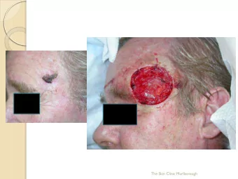
Treatment of skin injury • Major injury- can be Very Complex • Combined skills of • Wound care specialist • Dermatologist • Plastic surgeon and others • Best guidance: Refer patients to experienced providers with all information on radiogenic origin • Invariably experience may not be available, so take foreign help. Email…. Makes things easier. 33
Sequence • Dermatologist: Typically first to see • Dilemma: • He may not be aware • He is aware but patient does not know if the procedures he has undergone involves radiation, because interventionalist did not guide him • Diagnosis delayed for months 34
Cause of injury initially misidentified as pressure wound due to defibrillator pad. Injury ascribed to defibrillator pads- sued Lesion required grafting. company Grounding electrodes used for electrocautery 35
Consequences of misdiagnosis • Unnecessary dermatologic diagnostic procedures • Punch biopsy • Secondary complications 36
Ideal Situation- Diagnosis • Patient undergoes complex procedure • Skin dose > 5 Gy • Patient asked to keep watch and get back • Patient is called by hospital staff after 30 days • No chance of missing case, it will lead to correct diagnosis 37
38
Health Physics June 2010 (Vol.98, No.6) 39
40
Recommend


More recommend
Explore More Topics
Stay informed with curated content and fresh updates.