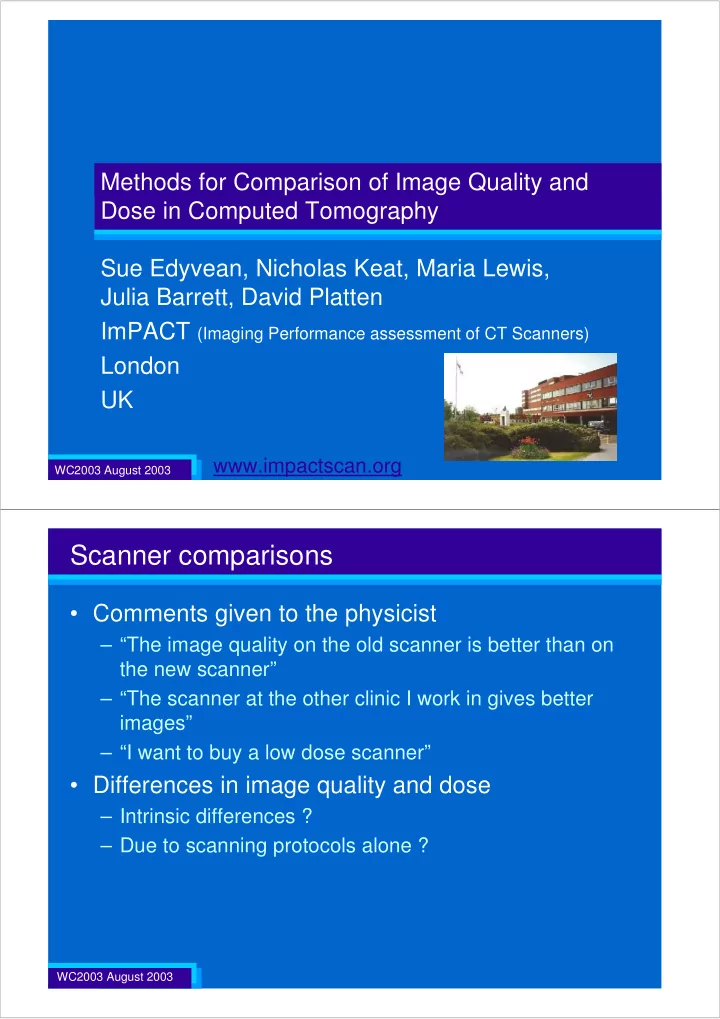

Methods for Comparison of Image Quality and Dose in Computed Tomography Sue Edyvean, Nicholas Keat, Maria Lewis, Julia Barrett, David Platten ImPACT (Imaging Performance assessment of CT Scanners) London UK www.impactscan.org WC2003 August 2003 Scanner comparisons • Comments given to the physicist – “The image quality on the old scanner is better than on the new scanner” – “The scanner at the other clinic I work in gives better images” – “I want to buy a low dose scanner” • Differences in image quality and dose – Intrinsic differences ? – Due to scanning protocols alone ? WC2003 August 2003
Image quality and dose dependence • Intrinsic factors ������ ���������� – Detectors x-ray tube slip rings • material • detector configuration • numbers of detectors – Data acquisition rates – Software corrections – Filtration – Focal spot x-ray detectors – Geometry • ie focus-axis, focus-detector distances WC2003 August 2003 Image quality and dose dependence • Scan protocols – Tube current – Tube voltage – Reconstruction algorithms – Collimation width – Helical pitch – Interpolation algorithms – Image slice thickness • Clinical application Image Plane WC2003 August 2003
Image quality and dose parameters • Comparison of image quality – Measured data not perception based information • Image quality – Image noise – Spatial resolution (scan plane) – Image thickness (spatial resolution in z-axis) • Radiation dose – CTDI vol WC2003 August 2003 Image Noise appropriate phantom image region of interest (roi): noise = standard deviation σ WC2003 August 2003
Spatial resolution (scan plane) high contrast edge ESF → LSF → MTF sharp image CT PTFE 100 smooth MTF (%) image 50 CT w 10 Position Along Edge spatial frequency (cm -1 ) WC2003 August 2003 Spatial resolution (scan plane)
Image thickness (z-axis spatial resolution) • Inclined Al ramps (axial): FWHM of z-sens profile WC2003 August 2003 Helical image thickness • images reconstructed at intervals of 1/10 of the slice thickness • mean CT number at centre used to create the profile 200 2.5mm 150 CT Numbers 100 50 tungsten or gold disc, 0.05 mm 0 -10 -8 -6 -4 -2 0 2 4 6 8 10 reconstructed image position mm WC2003 August 2003
Radiation Dose (CTDI vol ) • CTDI w = CTDI 100 averaged in scan plane = 1/3 .CTDI C + 2/3 .CTDI p • CTDI vol = CTDI w averaged along z-axis = CTDI w / Pitch Pitch 1 Pitch 2 WC2003 August 2003 Comparison of image quality and dose • Start with typical clinical protocol – (eg abdomen) minimise built in clinical application factors • Standardise scan parameters where possible – minimise effect from these variables – eg kV, pitch, collimation, nominal image width • Correct for known parameter inter-dependencies – eg. noise and dose, noise and image width • Obtain trend data for other inter-dependencies – eg. noise and resolution WC2003 August 2003
Scanning dependent factors • Noise with kV Noise Versus kV 2.00 Hard Beam Scanner A Values Soft Beam Scanner B 1.50 normalised to 120 kV 1.00 0.50 80 100 120 140 kV WC2003 August 2003 Scanning dependent factors Relative CTDI 2.4 2.2 2.0 relative CTDI four and sixteen slice 1.8 poor single slice 1.6 good single slice 1.4 1.2 1.0 0.8 0 5 10 15 20 25 30 35 collimation WC2003 August 2003
Standardise scan parameters • Single slice scanner • Nominal image width = collimation width • Multi slice scanner – Axial • Collimation width (eg. 10 mm) • Detector acquisition width (eg 2.5 mm) = nominal image width – Spiral • Collimation width (e.g 10 mm) • Detector acquisition width (eg 2.5 mm) • Nominal reconstructed Image width z-axis eg 10 mm collimation: 4 x 2.5 mm WC2003 August 2003 Correct for known parameter inter-dependencies • Noise with dose (tube current), slice thickness 2 1 2 1 ( ) ( ) ( ) ( ) α α noise α α noise α α α α mA slice thickness WC2003 August 2003
Identify other inter-dependent relationships • Noise, resolution ← reconstruction filter, focal spot • Noise and resolution with reconstruction filter – GE Lightspeed h/b: soft, standard, lung, detail, bone, edge – Siemens Volume Zoom h/b: AH/AB..10,20,30,40,50,60,70 – Toshiba Aquilion h: FC20,21,22,23,24,25,26,27,28,30,80: b: FC….10,11,12,13,14,30 – Marconi (Philips) MX8000 h: A,EB,EC,B,C,D; b: A,EC,B,C,D WC2003 August 2003 Noise and reconstruction filter 10.0 %noise for 40 mGy and 5 mm image width noise and resolution images reconstructed Siemens S4 with different algorithms % noise for 40 mGy 1.0 0.1 3.0 4.0 5.0 6.0 7.0 8.0 9.0 10.0 11.0 Average (MTF50, MTF10) WC2003 August 2003
Noise and reconstruction filter 10.0 %noise for 40 mGy and 5 mm image width GE LightSpeed Plus A Philips Mx8000 B Siemens S4 C D Toshiba Aquilion % noise for 40 mGy 1.0 0.1 3.0 4.0 5.0 6.0 7.0 8.0 9.0 10.0 11.0 Average (MTF50, MTF10) WC2003 August 2003 Noise and reconstruction filter Power fits 0.70 R 2 = 0.9998 GE LightSpeed Plus A R 2 = 0.9491 Philips Mx8000 0.65 B R 2 = 0.9969 Siemens S4 C R 2 = 0.2977 D Toshiba Aquilion 0.60 0.55 % noise for 40 mGy 0.50 0.45 0.40 0.35 0.30 0.25 0.20 3.0 3.5 4.0 4.5 5.0 5.5 6.0 6.5 Average (MTF50, MTF10) WC2003 August 2003
Noise and reconstruction filter 0.70 Linear fits R 2 = 0.9996 GE LightSpeed Plus A R 2 = 0.9585 0.65 Philips Mx8000 B R 2 = 0.9823 Siemens S4 C R 2 = 0.2833 0.60 Toshiba Aquilion D 0.55 % noise for 40 mGy 0.50 0.45 0.40 0.35 0.30 0.25 0.20 3.0 3.5 4.0 4.5 5.0 5.5 6.0 6.5 Average (MTF50, MTF10) WC2003 August 2003 Image quality and dose: normalise data Noise Resolution Image width Dose A 3 HU 3.4 c/cm 5 mm 20 mGy B 4.5 HU 3.8 c/cm 4.5 mm 25 mGy A 3 HU 3.4 c/cm 5 mm 20 mGy B 3 HU 3.4 c/cm 5 mm 36 mGy WC2003 August 2003
Image quality and dose: normalise data Noise Resolution Slice width Dose A 3 HU 3.4 c/cm 5 mm 20 mGy B 4.5 HU 3.8 c/cm 4.5 mm 25 mGy A 3 HU 3.4 c/cm 5 mm 20 mGy B 4 HU 3.4 c/cm 5 mm 20 mGy WC2003 August 2003 Image quality and dose: display data 10.0 %noise for 40 mGy and 5 mm image width GE LightSpeed Plus A Philips Mx8000 B Siemens S4 C D Toshiba Aquilion % noise for 40 mGy 1.0 0.1 3.0 4.0 5.0 6.0 7.0 8.0 9.0 10.0 11.0 Average (MTF50, MTF10) WC2003 August 2003
Image quality and dose: single numerical factor • Noise, dose and slice thickness relationship – established 1 1 2 α 2 α σ σ z D • Noise and resolution relationship – from graph (best fit) – theoretical WC2003 August 2003 Theoretical : imaging theory • Rodney Brookes and Giovanni di-Chiro (1976) – Statistical limitations in x-ray reconstructive tomography • Medical Physics Vol 3, No 4 July 1976 • Riederer S.J., Pelc N.J. and Chesler D.A. (1978) – The Noise Power Spectrum in Computed Tomography • Physics in Medicine and Biology 1978 23(3), 446-454 limiting resolution 1 2 α σ f 3 z 2 α 3 / 2 f σ α σ z D 1 2 α σ D WC2003 August 2003
Image quality and dose • Single numerical quality factor 3 / 2 f 3 f Q const . 2 α = σ z D σ z D • High Q – High image quality • high resolution, low noise and thin slice – Low dose WC2003 August 2003 Image quality and dose: single numerical factor Noise Resolution Slice width Dose A 3 HU 3.4 c/cm 5 mm 20 mGy Q = 1.0 Q = 0.7 B 4.5 HU 3.8 c/cm 4.5 mm 25 mGy A 3 HU 3.4 c/cm 5 mm 20 mGy B 3 HU 3.4 c/cm 5 mm 36 mGy WC2003 August 2003
Comparison of image quality and dose • Start with typical clinical protocol • Standardise scan parameters where possible – eg collimation, kV, pitch, nominal image width • Correct for known parameter inter-dependencies – eg. noise and dose, image width • Obtain trend data for other inter-dependencies – eg. noise and resolution • Review performance data – Normalise values to give single value of dose or noise – Display normalised noise against resolution – Calculate a single quality performance parameter WC2003 August 2003 Methods for Comparison of Image Quality and Dose in Computed Tomography Sue Edyvean, Nicholas Keat, Maria Lewis, Julia Barrett, David Platten ImPACT (Imaging Performance assessment of CT Scanners) London UK www.impactscan.org WC2003 August 2003
Recommend
More recommend