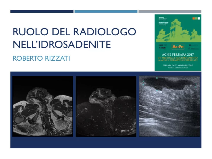

RUOLO DEL RADIOLOGO NELL’IDROSADENITE ROBERTO RIZZATI
DEFINITION Hidradenitis suppurativa (HS/AI): - Chronic - Inflammatory - Recurrent (at least 6 months) - Debilitating skin disease - Starting from the hair follicle - Usually presents after puberty - Painful, deep-seated, inflamed lesions - In the apocrine gland-bearing areas - Most commonly axillary, inguinal and anogenital regions
EPIDEMIOLOGY AND AETIOLOGY Mean age of onset: early ‘20 - It has also been reported in children and - postmenopausal women. Decline in prevalence after the age of 55 - Female / male ratio: 3-4/1 -
CLINICAL FEATURES ¡ Tipical lesions ¡ Tipical localizations Nodules Axillary ¡ ¡ Abscesses Inguinal ¡ ¡ Plaques Perianal ¡ ¡ Fistulae Gluteal ¡ ¡ Sinus tracts ¡ Scars ¡ ¡ Tipical evolution Slow progression ¡ Quick worsening ¡
Radiologo
WHAT TO ASK TO THE RADIOLOGIST? ¡ Differential diagnosis (Crohn’s disease) ¡ Staging ¡ Therapy response follow-up ¡ Complications assessment and follow-up
STAGING Hidradenitis suppurativa has 3 stages (Hurley stages): ¡ Solitary or multiple isolated abscess formation; no scarring or sinus tracts. Resembling acne. ¡ Recurrent abscesses, single or multiple widely separated lesions. Sinus tract formation is present. This can restrict movement and incision and drainage may be required. ¡ Diffuse or broad involvement across a regional area with multiple interconnected sinus tracts and abscesses. Fistulation and scarring.
CLINICAL CORRELATION AND STAGING
RADIOLOGY IN HIDROSADENITIS The diagnosis of hidradenitis suppurativa is clinical, and imaging is non-specific. ¡ Differential diagnosis ¡ Staging ¡ Follow-up
IMAGING • examination under anaesthesia (EUA) • pelvic magnetic resonance imaging (MRI) • anorectal endoscopic ultrasonography (EUS) • transcutaneous perianal ultrasound (TPUS) • fistulography and computed tomography (CT). US - MRI
ULTRASOUND ¡ A number of features can be identified by ultrasound. These features include both actual lesions and possible predisposing factors such as skin thickness and hair follicle morphology.
Sonografic Criteria of HS ¡ Widening of the hair follicles ¡ Thickening or abnormal echogenicity of the dermis ¡ Dermal pseudocystic nodules (round or oval-shaped hypoechoic or anechoic nodular structures) ¡ Fluid collections (anechoic or hypoechoic fluid deposits, in the dermis or hypodermis connected to the base of widened hair follicles) ¡ Fistulous tracts (anechoic or hypoechoic band-like structures across skin layers in the dermis or hypodermis connected to the base of widened hair follicles) Ultrasound In-Depth Characterization and Staging of Hidradenitis Suppurativa Wortsman et al Dermatologic Surgery October 2012
Right axilla of a 38-year-old female patient. Ultrasound findings of one of the lesions showing a hypoechogenic lesion showing vascularity at the periphery of the lesion; Kelekis et al british journal of dermatology february 2010
MRI (no mdc e.v!!) ¡ MRI is the test of choice to assess extent and for complications. MRI is also useful to differentiate from Crohn disease, the main differential diagnosis. ¡ STIR is considered the most useful sequence. ¡ marked thickening of the skin ¡ induration of the subcutaneous tissues ¡ formation of multiple subcutaneous abscesses ¡ prominent lymphadenopathy ¡ The differential diagnosis for these findings includes carbuncles, lymphadenitis, and infected Bartholin's or sebaceous cysts. Sinus and fistula formation remote from rectum and anus (cf. Crohn disease).
RADIOLOGICAL EVALUATION
Case 1 31-year-old woman with hidradenitis suppurativa
ANATOMY “Per capire le opzioni chirurgiche per il trattamento della malattia fistolosa, importante e conoscere l’anatomia e la funzione degli sfinteri”
CLASSIFICATION Parks classification of perianal fistula Intersfinterica (a) 45% ¡ Transfinterica (b) 30% ¡ Soprasfinterica (c) 20% ¡ Trans elevatore ano senza interessare gli sfinteri (d) 5% ¡ Fistola sottocutanea (e) (non inclusa nella class di Parks) ¡ St James’s University Hospital MR Imaging Classification of Perianal Fistulas RadioGraphics 2002
CASE 2 - MRI C.D. m 61 aa Tragitto fistoloso laterale sinistro con tragitto caudale che si dispone a “ferro di cavallo” in continuità con raccolta ascessuale glutea destra
CASE 2 - MRI O.Z. f 37 aa Tragitto fistoloso laterale sinistro ore 3 transfinterico con decorso in regione glutea mediale e orifizio cutaneo.
CASE 3 - US S.C. f 23 aa
CASE 3 - MRI S.C. f 23 aa
Conclusioni ¡ Staging ed eventuale planning pre operatorio ¡ Follow up complicanze o post terapia ¡ DD ¡ CEUS???? ¡ PDTA
Grazie per l’attenzione!! r.rizzati@ausl.fe.it
Recommend
More recommend