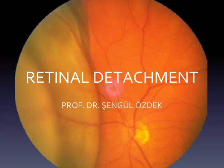

RETINAL DETACHMENT PROF. DR. ŞENGÜL ÖZDEK
Histoloji
Anatomy
RETINAL DETACHMENT • Separation of the neurosensory retina from retinal pigment epithelium. • Incidence 1 / 10.000, Risk is 3% until the age of 80 • Bilaterality 10% • Most common: 40-70 year-old
TYPES • RHEGMATOGENOUS RD • TRACTIONAL RD (PDR, VENOUS OCCLUSIVE DISEASE…) • EXUDATIVE RD (ECLAMPSIA, KMM)
The powers holding retina in place • Vitreous pressure • Passive fluid flow from vitreous to choroid • RPE tight junctions • RPE active ion transport • Bruch membrane (flow from RPE to choroid) • Concentration gradients (ionic, osmotic)
RRD D evelops in three stages • Posterior vitreous detachment • Retinal break / tear • Retinal detachment
Posterior Vitreous Detachment Stronger adhesions: • Vitreous base • Around the optic nerve head • Macula • Retinal big vessels • Around the retinal degenerations areas
ACUTE PVD A fter development of synchisis in some persons, small breaks occur in posterior vitreous cortex and liquefied vitreous passes to retrohyaloid space
ACUTE PVD • The remaining solid vitreous collapse down and retrohyaloid space filled with sinchitic fluid: PVD • Sensorial retina lacks protection • Sensorial retina is vulnerable to vitreoretinal traction
PVD • More in elderly, myopics, aphakic / pseudophakic patient and people exposed to trauma • Mostly asymptomatic • Photopsia (flashes of light) • Gliotic tissue which adheres to the posterior hyaloid membrane where papilla and vitreous opacities: Floaters (flight of fly)
Acute PVD Complications • Retinal Tear • Macular Hole • Epiretinal Membrane
Acute PVD’s Complications • Vessel avulsion • Vitreous hemorrhage
Peripheral retinal degenerations
Lattice degeneration (lattice = wire netting) • Most important peripheral degeneration • It is a band-shaped retinal thinning, in front of the equator, parallel to the ora serrata, which contains lines in the form of wire netting. • atrophy starts from the inner limiting membrane and spreads to the other lines • In the middle of degeneration vitreous is liquefied but at the edge of degeneration vitreous is attached
Retinal break Horseshoe tears Holes Disinsertion ( dialysis )
HORSE-SHOE TEAR The most common reason for RD • The apex located toward to central • Photopsia + Floaters + • If accompanied by the rupture of blood vessels: blurred vision
Retinal Holes • Asymptomatic • Within lattice dehgeneration areas • Punched out circular holes
Mechanism of RD
DISINSERTION (DIALYSIS) • In severe blunt trauma • Usually in inferior temporal quadrant • Severe photopsia • Detachment may not occur for many years in young patient if vitreous can remain gel formation
PVR • Proliferative Vitreoretinopathy (PVR) • The proliferation of RPE cells and gliotic cells • Long term RD • Giant and a multible number of breaks • Penetrating injury • Vitreous hemorrhage • Fast wound healers
PVR Stages Grade A : Vitreous haze, pigment clumbs in vitreous and inferior surface of the retina ( tobacco dust ) Grade B : creases on the face of inner retina, decreased mobility of vitreous gel and retina, irregular tear edges, tortuosity of blood vessels Grade CP: behind equator local, diffuse or peripheral retinal creases, subretinal cords Grade CA: Same appearance in front equator and cords in condensed vitreous
Myopia - RD • 10% of the general population: Myopic • 40% of all RDs occur in myopic eyes. • Lattice deg. is more common in -6.0 -9.0 myopes • Vitreous degeneration and PVD are more common in myopes
Trauma - RD • 10% of RD occurs following trauma. • The most common cause of RD in children • Severe blunt trauma: retinal dialysis, macular hole • Penetrating injury: Both tractional and RRD.
RD Symptoms • The first sings of acute PVD are fotopsia and floaters • Peripheral visual field defect: like a black curtain one side of the eye • After macula is affected, VA will decrease to hand motions only
RRD signs • IOP: 5 mmHg lower • Retinal break • Detached Retina has a convex configuration and an opaque appearance
Treatment • PROPHYLAXIS IS VERY IMPORTANT – Acute PVD’s Symptoms: Photopsia, floaters: peripheral retinal examination! – Myopia or trauma or family history or fellow eye history of RD: detailed fundus examination! – Symptomatic or dangerous peripheral retinal degenerations and retinal tears: laser
Retinal Detachment Surgery 1. External buckling: Peripheral or local scleral buckling: Classic Technique 2. İnternal retinopexy: PPV-tamponade – laser or cryo to tears – Gas-Silicone oil
Scleral Buckle • Silicone band or with local sponge • Intraoperative cryotherapy around the tear • Drainage of Subretinal fluid. • IV Air-Gas
Internal retinopexy: Tamponade • Gas: SF6, C3F8 • Air
PPV • Associated Vitreous Hemorrhage, • PVR, • Multible/giant tears • Macular holes
Tractional RD 1. PDR: Proliferative diabetic retinopathy 2. ROP prematurity of retinopathy 3. Penetrating trauma 4. Sickle cell anemia, Vein occlusions, PFV • Retina is immobile, surface is concave. • Tractions may cause tears... COMBINED FORM RD
Traksiyonel RD
Trauma
PFV
ROP
ROP Stage 5: Total RD-Leukocoria
Tractional RD • Photopsia and floaters (-) • Vision loss occurs slowly • Treatment: PPV
Exudative RD • Malign hypertension • Hypertensive crisis/Eclampsia • Vascular: Coats desease • Tm: CMM, Metastases, choroidal hemangioma • Uveitis: Vogt-Kayanagi- Harada • Central serous chorioretinopathy
Exudative RD • Exudative RD: fluid leaks from retinal vessels and RPE • there is no tear and traction. • May move with gravity and head movements
Exudative RD • Vision is very low in the morning due to the liquid which reason to detachment becomes the subject of gravity. When patient seats, vision begins to improve.
SSKR
Exudative RD • No Photopsia, • Floaters (+/-): becauase of vitritis • Visual field defect suddenly • No surgical treatment. • Treatment of the underlying condition.
Recommend
More recommend