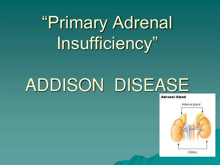

“Primary Adrenal Insufficiency” ADDISON DISEASE
Adrenocortical insufficiency comprises of primary and secondary adrenal insufficiency. Primary adrenal insufficiency can be congenital or acquired. Acquired primary adrenal insufficiency is termed as Addison disease.
Addison Disease is the result of an under active adrenal gland. An under active adrenal gland produces insufficient amounts of cortisol (hormones that help control the body's use of fats, proteins and carbohydrates, suppress inflammatory reactions in the body, and affect immune system function) and Aldosterone (that maintains body salts and water).
Pathophysiology liver function low sugar Cortisol Very Low digestive enzyme vomiting, diarrhea Non functioning Adrenal gland Brain, Coma kidney, Na Low fluid Aldosterone & water loss volume very Low Shock . Heart, irregular Low BP Decrease out put
ETIOLOGY Autoimmune adrenalitis – Isolated autoimmune adrenalitis – Autoimmune adrenalitis as a part of APS APS type 1 APS type 2 APS type 4 Infectious Adrenalitis – Tuberculous adrenalitis – Fungal adrenalitis – HIV associated Bilateral Adrenal Haemorrhage Bilateral Adrenalectomy
Genetic Disorders – Adrenoleukodystrophy – Congenital adrenal hyperplasia – Congenital lipoid adrenal hypoplasia – ACTH insensitivity syndrome – Smith-Lemli-Opitz syndrome – Triple A syndrome Adrenal infiltration Drug induced adrenal insufficiency
Autoimmune Addison Disease Autoimmune destruction of the adrenal glands is the most common cause of Addison Disease. Component of autoimmune polyendocrinopathy syndromes: • APS type 1: Chronic mucocutaneous candidiasis Hypoparathyroidism Addison Disease • APS type 2: Addison disease with Autoimmune thyroid disease (Schmidt syndrome) or Type 1 Diabetes Mellitus (Carpenter Syndrome)
Infections Most frequent infectious etiology is Meningococcemia. Tuberculosis is Second common cause of adrenal destruction. HIV infection.
Drugs Ketoconazole Rifampicin Phenobarbitone Phenytoin Mitotane
Haemorrhage into Adrenal Gland Breech presentation Anticoagulant therapy Child Abuse
HOW THEY PRESENT
Hypoglycemia (sweating and irritability) Hypotension Hypovolemia Hyponatremia Hyperkalemia
Hyperpigmentation Marked at exposed areas, buccal and gingival mucosa, at scars and genitalia.
Non specific signs: • Muscle weakness • Malaise • Anorexia • Weight loss • Orthostatic hypotension
HOW TO INVESTIGATE
Serum electrolytes: – Hypoglycemia – Hyponatremia – Hyperkalemia ABG Renal function Plasma renin activity Urinary Sodium, and potassium levels CT scan and MRI
Specific: Serum cortisol levels (low) Before stimulation After stimulation i.e. 30 – 60 min after administration of 0.25mg cosyntropin
MANAGEMENT
IMMEDIATE MANAGEMENT • Intravenous administration of 5% dextrose. • 0.9% saline solution • Hydrocortisone • Hydrocortisone sodium succinate • Intravenously, 6 hourly for 24 hours • 10mg in infants, 25mg in toddlers,50 mg in children, and 100 mg in adolescent. • Treat hyperkalemia
LONG TERM MANAGEMENT • Cortisole replacement • Hydrocortisone • Daily dose: • 10 mg/m2/24 hours • 3 divided doses • High doses: • 2 – 3 folds increased dose • Infection • Stress • Minor surgery • Major surgery (intravenous steroids) • Aldosterone replacement • Fludrocortisone • 0.05 – 0.3 mg per day orally
Follow up • Adherence to treatment • ACTH levels are main stay to monitor the adequacy of replacement therapy • Adverse effects of treatment • Frequency of crisis
Recommend
More recommend