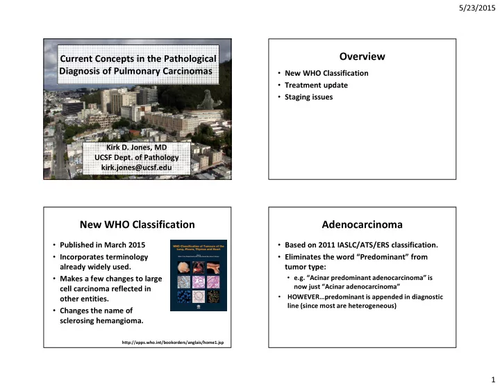

5/23/2015 Overview Current Concepts in the Pathological Diagnosis of Pulmonary Carcinomas • New WHO Classification • Treatment update • Staging issues Kirk D. Jones, MD UCSF Dept. of Pathology kirk.jones@ucsf.edu New WHO Classification Adenocarcinoma • Published in March 2015 • Based on 2011 IASLC/ATS/ERS classification. • Incorporates terminology • Eliminates the word “Predominant” from already widely used. tumor type: • e.g. “Acinar predominant adenocarcinoma” is • Makes a few changes to large now just “Acinar adenocarcinoma” cell carcinoma reflected in • HOWEVER…predominant is appended in diagnostic other entities. line (since most are heterogeneous) • Changes the name of sclerosing hemangioma. http://apps.who.int/bookorders/anglais/home1.jsp 1
5/23/2015 Adenocarcinoma Types • Lepidic • Acinar • Papillary • Solid • Micropapillary • These are the major non-mucinous types Lepidic: Surface alveolar growth of tumor cells Acinar: Round, oval, or irregular glands invading in a fibrous stroma 2
5/23/2015 Cribriform pattern included in acinar Papillary: Tumor cells grow on surface of fibrovascular cores Solid: Sheets of tumor cells without gland formation 3
5/23/2015 Solid without mucin production – former large cell Micropapillary: Growth as small papillae without fibrovascular cores Other Adenocarcinomas • Invasive mucinous adenocarcinoma • Colloid adenocarcinoma • Fetal adenocarcinoma • Cribriform pattern (currently under acinar, but behaves like solid) 4
5/23/2015 Invasive mucinous adenocarcinoma: Often shows lepidic growth… Invasive mucinous adenocarcinoma: …admixed with acinar pattern Colloid adenocarcinoma: Abundant pools of mucin replacing alveoli… Colloid adenocarcinoma: …with tumor cells floating as clusters and alveolar walls. 5
5/23/2015 Architecture as Grade • Lepidic = Grade 1 • Acinar and Papillary = Grade 2 • Solid and Micropapillary = Grade 3 • Mucinous, colloid, fetal = Grade 3 • There is an additional grading scheme using nuclear grade and mitoses that helps divide Yoshizawa A, et al. Mod Pathol. 2011 May;24(5):653-64. the 2’s von der Thüsen JH, et al. J Thorac Oncol. 2013 Jan;8(1):37-44. Prognosis by Pattern • Micropapillary type shows worse prognosis. • Zhang J, et al. Histopathology. 2011 Dec;59(6):1204-14 Yoshizawa A, et al. Mod Pathol. 2011May; 24(5): 653-64. 6
5/23/2015 Any Micropapillary? Any Micropapillary? Central scar tissue (red), Acinar (yellow), Papillary (blue), and Micropapillary (green). Lee G, et al. Am J Surg Pathol. 2015 May;39(5):660-6. PMID: 25724001. Lee G, et al. Am J Surg Pathol. 2015 May;39(5):660-6. PMID: 25724001. Semiquantitative Analysis Adenocarcinoma Variants • Divide into patterns based on 5% increments. • Does it matter to the clinician? Then divide into predominant pattern. • What to put on the bottom line • “Weak recommendation, low-quality - Adenocarcinoma with a comment. evidence” - ____-predominant adenocarcinoma. • I mention if micropapillary pattern is present. • Lepidic pattern (AIS) has the same clinical intrigue as BAC used to have. 7
5/23/2015 Spread through air spaces (STAS) Judging Invasion • Micropapillary clusters, solid nests, or single • Concept of AIS and MIA cells present within alveoli outside of the • Clear invasion main tumor mass. • pattern that is not lepidic • Likely result in cases of localized recurrence • vascular or pleural invasion after limited resections. • STAS • Mention if present, particularly if present at • fibromyxoid stroma (desmoplasia) margin (margin is negative for invasive tumor, but presence of STAS correlated with increased risk of local recurrence). Judging Invasion Squamous Cell Carcinoma • Difficult to judge collapse of lepidic growth • Previously defined histologically by from acinar pattern keratinization • Difficult to judge collapse of lepidic growth • Now two types: from papillary • Keratinizing • Some choose to just measure the region of • Non-keratinizing collapse (Noguchi B type) • Proper to measure the limited area of fibromyxoid tissue - difficult 8
5/23/2015 p40 9
5/23/2015 Potential Pitfalls Large Cell Carcinoma • TTF-1: • Previously used when no morphologic – Thyroid carcinoma support for squamous cell or adenocarcinoma – Entrapped pneumocytes • Now use immunohistochemical stains to help – Gyn tumors (~80% ut. carcinosarcoma) subclassify into: – Neuroendocrine tumors • Solid type adenocarcinoma • Napsin-A: • Non-keratinizing squamous cell carcinoma – Pulmonary macrophages (darker) • Large cell carcinoma – Renal cell carcinoma (~80%) – GI mucinous tumors (~80%) Mystery Case Potential Pitfalls • p63: • 64-year-old woman with right lower lobe – Entrapped basal layer lung nodule. – Urothelial tumors • CT-guided percutaneous fine needle – Metastatic squamous tumors aspiration performed. – Adenocarcinoma of lung • Require >10% of nuclei to stain • p40: – More specific, but similar pitfalls 10
5/23/2015 Using the CT scan Bone tumors, ILD, and now lung tumors • Ground glass opacities versus solid masses - Determining extent of lepidic growth - Determining size of lesion • Border of a lesion - Spiculated versus smooth - Typical adeno vs benign or fast-growing • Multiplicity of lesions - Extrathoracic with met, lung met, synch primary 11
5/23/2015 The Argus (Melbourne, Australia) February 6, 1936, page 10 Sclerosing Pneumocytoma • Formerly sclerosing hemangioma – “Sclerosing hemangioma (histiocytoma, xanthoma) of the lung” – A.A. Liebow and D.S. Hubbell, Cancer, 1953. • Characteristic radiologic appearance – Rounded edges are often either really bad (fast growing) or benign • Characteristic immunoprofile – EMA positive, Keratin negative – TTF-1 positive, Napsin-A negative • Immature pneumocytes with surface normal bronchiolar epithelium Keratin 12
5/23/2015 EMA Napsin-A Treatment Options • Many tumors are typically treated with standard chemotherapy • In recurrent and stage 4 tumors, and increasingly as first line, targeted treatments being used: • EGFR • BRAF • EML4-ALK • MET • ROS-1 52 TTF-1 13
5/23/2015 Resistance Mutations Immunotherapy • EGFR TKI-treated tumors often develop • PD-1 and PD-L1 additional mutations – Programmed death 1 receptor and its ligands – commonly T790M within EGFR – PD-1 is an inhibitory checkpoint pathway in T cells – Novel TKI – Some tumor cells have increased surface expression of PD-L1 (35-95% of NSCLC) • Targeting other pathways being activated – Currently in trials (although already FDA approved), – MET, AXL most often for patients that have failed first and second line therapies Staging Issues Multiple Nodules • Multiple nodules • Sometimes difficult to determine if two tumor nodules represent • Pleural invasion – Synchronous primary tumors • Pleural drop metastases – Intraparenchymal metastases 14
5/23/2015 Comprehensive Histologic Martini-Melamed Assessment • Tumors are synchronous primaries if: • The “histologically different” component is 1. Histologically different. expanded substantially 2. Histologically similar but… – Percentage of adenocarcinoma subtype A. Arise from CIS becomes significant B. No tumor in shared lymphatics – Cytologic features, stromal components also aid differentiation C. No extrapulmonary mets • Additional concept of AIS/Lepidic growth • At the time, histologically different meant SqC vs adeno, and CIS was Sq.CIS • To be discussed in the new AJCC – next year? Pleural invasion • The many definitions of pleural invasion – What we want to think versus what there is data to support – Research from Japan (lots more EVG staining going on overseas) • The prominent elastic layer (the visceral pleural elastica, aka the external elastic layer) 15
5/23/2015 16
5/23/2015 17
5/23/2015 Pleural Invasion Pleural Drop Metastases • EVG for all tumors approaching the pleura. • Tumor studding on pleural surface • pT2a if external elastic layer is penetrated • NOT direct extension (T2, PL2) (visceral pleural elastica). • NOT subpleural lymphatic invasion with – Raises stage from IA to IB in small tumors. • Elastica of chest wall is variable, and it is spread to other areas of the lung (T3 or T4) sometimes difficult to assess chest wall • Similar prognosis as malignant pleural invasion. effusion (M1a) – Look for penetration into parietal fat. • Can use PL designations if desired – Past elastica PL1, on pleural surface PL2, into chest wall PL3 18
5/23/2015 Take Home Messages • No significant changes to adenocarcinoma terminology since IASLC/ATS/ERS changes. • Splitting of large cell using IHC. • Sclerosing pneumocytoma. • Targeted therapy, targeting resistance, immunotherapy. • Not all multiple lesions mean poor prognosis. • Treat the pleura with respect. 19
Recommend
More recommend