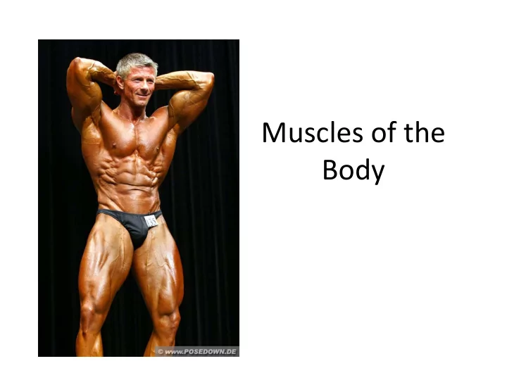

Muscles of the Body
Muscles of the Body I. Movement and Leverage Systems II. Muscles action III. Fascicle Arrangement IV. Criteria used in naming muscles V. The Muscles (Lab) 2
I. Movement & Leverage Systems A. What is and Why use levers? 1. What it is: a. Physics definition: a rigid bar that pivots about one point and is used to move an object at a second point by a force applied at a third. b. Altered for anatomy: rigid bars (bones) that pivot about a point (joint) and is used to move an object at a second point by a force applied (muscles) at a different point (this point depends on the lever classification). 2. Why use: a. to gain a mechanical advantage as it relates to movement of the body… usually in speed and strength 3
I. Movement & Leverage Systems B. Leverage components 1. Leverage systems require at least three components a. Lever – a rigid bar that moves (bones) b. Fulcrum – a fixed point (joint) c. Effort – applied force (muscles) d. Load – resistance (weight/mass) C. Classification of levers 1. First Class 2. Second Class 3. Third Class 4
Lever Systems – Mechanical Advantage Example 5
I. Movement & Leverage Systems C. Classification of Lever Systems 1. First ‐ class lever Effort applied at one end • Load is at the opposite end • Fulcrum is located between load and effort • Example? • 6
I. Movement & Leverage Systems C. Classification of Lever Systems 1. Second ‐ class lever Effort applied at one end • Fulcrum is at the opposite end • Load is between the effort and • fulcrum An uncommon type of lever in the • body Work at a mechanical advantage • • Example? 7
I. Movement & Leverage Systems C. Classification of Lever Systems 3. Third ‐ class lever • Effort is applied between the load and the fulcrum Work speedily • Always at a mechanical disadvantage (why?) • Example? • 8
II. Muscle Actions A. General Principles of Muscle Action 1. A muscle cannot reverse the movement it produces a. Remember: muscle tissue when active ONLY shortens! 2. Another muscle must undo the action 3. Muscles with opposite actions typically lie on opposite sides of a joint (if not, the force is directed to the opposite side via tendon). 9
II. Muscle Action A. Muscle Action Terms: 1. Prime mover (agonist) has major responsibility for a certain movement • Example: • 2. Antagonist opposes or reverses a movement • Example: • 3. Synergist helps the prime mover by: • By adding extra force – By reducing undesirable movements – Example: • 4. Fixator a type of synergist that holds a bone firmly in place • Example: • 10
III. Arrangement of Fascicles A. General Info to remember: 1. Skeletal muscles – consist of fascicles 2. Fascicles – arranged in different patterns 3. Fascicle arrangement – tells about action of a muscle 11
III. Arrangement of Fascicles B. Types of fascicle arrangement 1. Parallel – fascicles run parallel to the long axis of the muscle a. Strap ‐ like – sternocleidomastoid b. Fusiform – biceps brachii 2. Convergent a. Origin of the muscle is broad b. Fascicles converge toward the tendon of insertion c. Example – pectoralis major 12
III. Arrangement of Fascicles B. Types of fascicle arrangement cont… 3. Pennate a. Unipennate – fascicles insert into one side of the tendon b. Bipennate – fascicles insert into the tendon from both sides c. Multipennate – fascicles insert into one large tendon from all sides 4. Circular – fascicles are arranged in concentric rings a. Surround external body openings b. Sphincter – general name for a circular muscle c. Examples – orbicularis oris and orbicularis oculi 13
III. Arrangement of Fascicles 14
IV. Naming the Skeletal Muscles A. Location Example – the brachialis is located on the arm – B. Shape – Example – the deltoid is triangular C. Relative size Maximus, minimus, and longus indicate size – Example – gluteus maximus and gluteus minimus – 15
IV. Naming the Skeletal Muscles D. Direction of fascicles and muscle fibers Name tells direction in which fibers run – Example – rectus abdominis or circularis – oculi E. Location of attachments – name reveals point of origin and insertion Example – brachioradialis or – sternocleidomastoid 16
IV. Naming the Skeletal Muscles F. Number of origins – two, three, or four origins Indicated by the words biceps, triceps, and – quadriceps G. Action – the action is part of the muscle’s name Indicates type of muscle movement – Flexor, extensor, adductor, or abductor • 17
V. Muscles 18
Superficial Muscles of the Body – Anterior View 19
Superficial Muscles of the Body – Posterior View 20
Muscles of the Head Facial Expression • Muscles of facial expression • Lie in the face and scalp • Thin and variable in shape • Often insert in the skin – not on bones • Innervated by cranial nerve VII – the facial nerve 21
Muscles of the Head Facial Expression 22
Muscles of the Head – Mastication and Tongue Movement • Four main pairs of muscles involved in mastication – Innervated by mandibular division – the trigeminal nerve – Prime movers of jaw closure – masseter and temporalis – Side ‐ to ‐ side movement – pterygoid muscles – Compression of cheeks – buccinator muscles 23
Muscles of the Head Mastication and Tongue Movement 24
Muscles of the Head Mastication and Tongue Movement • Extrinsic muscles of the tongue • Move tongue: – Laterally – Anteriorly – Posteriorly • All innervated by cranial nerve XII – the hypoglossal nerve 25
Muscles of the Head Mastication and Tongue Movement 26
Muscles of the Anterior Neck and Throat Swallowing • The neck is divided into anterior and posterior triangles • Anterior triangle – Divided into suprahyoid and infrahyoid muscles – Participate in swallowing • Pharyngeal constrictors – squeeze food into the esophagus 27
Muscles of the Anterior Neck and Throat Swallowing 28
Muscles of the Anterior Neck and Throat Swallowing 29
Muscles of the Neck and Vertebral Column • Head movement – Sternocleidomastoid – Splenius capitis and splenius cervicis 30
Muscles of the Neck and Vertebral Column • Trunk extension – Deep muscles of the back • Maintain normal curvatures of the spine • Form a column from sacrum to the skull – Erector spinae group – largest of the deep back muscles 31
Muscles of the Neck and Vertebral Column 32
Deep Muscles of the Thorax – Breathing • Deep muscles provide movements for breathing – External intercostal muscles • Lift the ribcage – Internal intercostal muscles • May aid expiration during heavy breathing • Diaphragm – most important muscle of respiration – Flattens as it contracts – increases the volume of the thoracic cavity 33
Deep Muscles of the Thorax – Breathing 34 Figure 11.10a
Deep Muscles of the Thorax – Breathing 35
Muscles of the Abdominal Wall • Lateral and anterior abdominal wall – Formed from three flat muscle sheets • External oblique • Internal oblique • Transversus abdominis – Fourth muscle pair • Rectus abdominis – inserts at linea alba 36
Muscles of the Abdominal Wall 37
Muscles of the Pelvic Floor • Pelvic floor (pelvic diaphragm) • Sheet of two muscles – both support pelvic organs – Levator ani – formed from iliococcygeus, puborectalis, and pubococcygeus – Coccygeus 38
Muscles of the Pelvic Floor 39
Muscles of the Perineum • Inferior to the muscles of the pelvic floor • Urogenital diaphragm formed from: – Sphincter urethrae and the deep transverse perineus 40 Figure 11.12b
Muscles of the Perineum • Muscles of the superficial perineal space – Ischiocavernosus – Bulbospongiosus – Superficial transverse perineus 41
Superficial Muscles of the Anterior Thorax • Movements of the scapula – Pectoralis major – Pectoralis minor – Serratus anterior – Subclavius 42
Superficial Muscles of the Posterior Thorax • Movements of the scapula – Trapezius – Levator scapulae – Rhomboid major – Rhomboid minor 43
Muscles Crossing the Shoulder Joint • Movements of the arm – Deltoid – Pectoralis major 44
Muscles Crossing the Shoulder Joint • Movements of the arm – Latissimus dorsi – Supraspinatus – Infraspinatus – Teres minor – Teres major – Coracobrachialis – Subscapularis 45
Muscles Crossing the Elbow Joint • Posterior muscles – extensors of the forearm – Triceps brachii – Anconeus • Anterior muscles – flexors of the forearm – Biceps brachii – also supinates the forearm – Brachialis – Brachioradialis 46
Muscles of the Forearm • Movements of the wrist, hand, and fingers – Tendons are anchored by • Flexor and extensor retinacula – Most forearm muscles arise from the distal humerus – Movements at the wrist include: • Flexion, extension, abduction, and adduction – Wrist and fingers are “operated” by muscles in the forearm 47
Recommend
More recommend