MRI in Myocarditis MRI in Myocarditis Faculty of Medicine, - PowerPoint PPT Presentation
XVth Balkan Congress of Radiology XVth Balkan Congress of Radiology Danubius Hotel Helia, 12-14 October 2017, Budapest, Hungary Danubius Hotel Helia, 12-14 October 2017, Budapest, Hungary Ruica Maksimovi Ruica Maksimovi MRI in
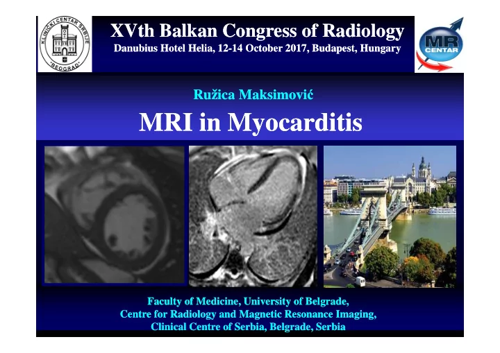
XVth Balkan Congress of Radiology XVth Balkan Congress of Radiology Danubius Hotel Helia, 12-14 October 2017, Budapest, Hungary Danubius Hotel Helia, 12-14 October 2017, Budapest, Hungary Ružica Maksimovi ć Ružica Maksimovi ć MRI in Myocarditis MRI in Myocarditis Faculty of Medicine, University of Belgrade, Faculty of Medicine, University of Belgrade, Centre for Radiology and Magnetic Resonance Imaging, Centre for Radiology and Magnetic Resonance Imaging, Clinical Centre of Serbia, Belgrade, Serbia Clinical Centre of Serbia, Belgrade, Serbia
Dg & Treatment of Myocarditis: ESC Consensus 2013 Definition of Inflammatory Cardiomyopathy Caforio, Pankuweit et al. Eur Heart J 2013 � Myocarditis in association with cardiac dysfunction (involved in the pathogenesis of DCM) � Idiopathic � Autoimmune � Infectious subtypes � WHF/ISFC expert pannel in 1997 set the immunhistochemical criteria: Minimum of 14 activated lymphocytes/mm 2 is neccessary for the diagnosis (diffuse or focal infiltrates with or without signs of hypertrophy or fibrosis � Viral cardiomyopathy: viral persistence in a dilated heart � Inflammatory viral cardiomyopathy (or viral myocarditis with cardiomegaly)
Etiology of Myocarditis Caforio, Pankuweit et al. Eur Heart J 2013
Development of Myocarditis Kawai C. From myocarditis to cardiomyopathy: mechanisms of inflammation and cell death: learning from the past for the future. Circulation 1999;99:1091–100.
Dg & Treatment of Myocarditis: ESC Consensus 201 3 Diagnostic Criteria for Suspected Myocarditis Caforio, Pankuweit et al. Eur Heart J 2013 Clinical Presentations: � Acute chest pain, pericarditic or pseudo- ischaemic � New-onset (days up to 3 mths) or worsening of: dyspnoea at rest or exercise, and/or fatigue, with or without left and/or right heart failure signs � Subacute/Chronic (>3 months) or worsening of: dyspnoea at rest or exercise, and/or fatigue, with or without left and/or right heart failure signs � Palpitation, and/or unexplained arrhythmia symptoms and/or syncope, and/or aborted sudden cardiac death � Unexplained cardiogenic shock
Dg & Treatment of Myocarditis: ESC Consensus 2013 Diagnostic Criteria for Suspected Myocarditis Caforio, Pankuweit et al. Eur Heart J 2013 I ECG/Holter/stress test features Newly abnormal 12 lead ECG and/or I I I I I I I I V V V 1 V V V V V 1 1 1 1 1 1 1 Holter and/or stress testing: � II II II II V 2 V V V II II II II V V V V I to III ° AV, or bundle branch 2 2 2 2 2 2 2 block, � ST/T wave change V V 3 V V 3 III III III III III III III III V V V V � 3 3 3 3 3 3 sinus arrest � VT or VF and asystole, AF � aVR aVR aVR aVR aVR aVR aVR aVR V V V 4 V V V 4 V V 4 4 4 4 4 4 reduced R wave height, intraventricular conduction delay (widened QRS complex), aVL aVL aVL aVL V V V V V V V 5 V 5 aVL aVL aVL aVL 5 5 5 5 5 5 abnormal Q waves, � low voltage, frequent PVCs, V 6 V 6 V V V V V V aVF aVF aVF aVF aVF aVF aVF aVF 6 6 6 6 6 6 PSVT
Dg & Treatment of Myocarditis: ESC Consensus 2013 Diagnostic Criteria for Suspected Myocarditis Caforio, Pankuweit et al. Eur Heart J 2013 II. Myocardiocytolysis markers � Eevated TnT/TnI III. Functional and structural abnormalities on cardiac imaging (echo/angio/CMR) New, otherwise unexplained LV and/or RV structure and Before Treatment function abnormality: � regional wall motion or global systolic or diastolic function abnormality, � with or without ventricular dilatation, � with or without increased wall thickness, � with or without pericardial effusion, � with or without endocavitary thrombi. IV. Tissue characterisation by CMR � Oedema and/or LGE of myocarditic pattern After Treatment
Dg & Treatment of Myocarditis: ESC Consensus 2013 Diagnostic Criteria for Suspected Myocarditis Lake Louise Consensus Criteria Caforio, Pankuweit et al. Eur Heart J 2013 ≥ 2 criteria
Most Important Indications for CMR in Patients with Suspected Myocarditis Friedrich M. Circ Cardiovasc Imaging 2013:6:833. Kindermann I. J Am Coll Cardiol 2012;59:779.
Case Presentation – Biopsy Positive Myocarditis MRI LV EDD 57 mm RV EDD 24 mm LV ESD 36 mm EF 58%, EDV 149 mm, ESV 63 ml IVS 10 mm PW 10 mm EF 62%, EDV 159 mm, ESV 64 ml
Case Presentation – Biopsy Positive Myocarditis MRI Before CM After CM
Case Presentation Medical History � Male, 18 years, was examined due to dry cough, chest pain � Echocardiography revealed enlarged, hypocontractile LV LV EDD 80 mm LV ESD 74 mm LV EF 25% IVS 9 mm LVEDV 340 ml PW 9 mm LVESV 240 ml RV EDD 41 mm
Case Presentation Medical History
Case Presentation –Myopericarditis Medical History � Male, JS, 20 years � Echocardiography showed transient pericardial effusion with constrictive physiology, no regional wall motion Cine TrueFISP abnormalities of the LV � Elevated serum troponin � Chest x-ray, bilateral pleural effusion � Therapy: nonsteroidal anti-inflammatory drugs TSE T2w FS
Case Presentation –Myopericarditis MRI LVEF 66% LV EDD 53 mm LVEDV 151 mm LV ESD 31 mm LVESV 51 ml IVS 10 mm SV 100 ml PW 9 mm
Case Presentation Medical History � � Laboratory Male, 22 years admitted to the Troponin L 4 230 (0-0.04 ug/L) hospital due to suspected acute BNP 894 (0-73 pg/ml) coronary syndrom � C reactive protein 295.6 (0-8 mg/L) Had acute chest pain for an hour � Variations of Troponin L (April 22, Coronarography 7680, April 23, 10720, April 27, 4 Normal finding � 230) ECG � Treatment Sinus rhythm, elevation of ST Antiaggregation therapy segment Nonsteroid antrheumatic Blockers of H2 receptors and inhibitors of proton pump Electron Micrograph of a Parvovirus
Case Presentation MRI April 29, 2015 LV EDD 52 mm LV EF 35% LV ESD 33 mm LV EDV 180 ml IVS 9 mm LV ESV 117 ml PW 9 mm RV EDD 32 mm
Case Presentation MRI
Case Presentation MRI
Case Presentation MRI – Follow-up July 8, 2015 LV EDD 50 mm LV EF 64% LV ESD 38 mm LV EDV 145 ml IVS 9 mm LV ESV 117 ml PW 9 mm RV EDD 25 mm Recovery !
Case Presentation MRI – Follow-up
Case Presentation MRI – Myocarditis Before Contrast After Contrast
Case Presentation US – Peak Longitudinal Strain in Myocarditis X X
CMR Results in Relation to ECG and Troponin Florian A. Clin Res Cardiol 2015:104:154. ECG – ST elevation (I, V2-V6)
Clinical and MRI Parameters as Predictors of Outcome In Pediatric Myocarditis Sachdeva S. Am J Cardiol 2015:15:499. N-58 pts 16 year old male patient with chest pain, dyspnea and flu-like symptoms with troponin T elevation, ECG ST elevation V2 to V6. LGE showed extensive hyperenchancement with subepicardial distribution in the inferior, lateral and anterior wall and septum.
Summary of Recommended Components of The CMR Report for Myocarditis Friedrich M. JACC 2009:53:17. � LV end-diastolic volume and volume index LV volume and function � LV end-systolic volume and volume index � Ejection fraction � Cardiac index � LV mass and mass index � T2 signal/edema (regional edema or global T2 ratio) Presence or absence of markers for � Calculated global myocardial myocardial early inflammatory activity and injury of ST gadolinium enhancement ratio � Myocardial late gadolinium enhancement with non- ischemic regional distribution Based on the presence or absence of 2 or more criteria, Conclusion considering additional evidence by the presence of LV dysfunction and/or pericardial effusion Based on clinical setting Recommendation for follow up A follow-up >4 weeks after the onset of symptoms may have prognostic implications and thus is recommended.
Conclusions M. Friedrich. Circ Cardiovasc Imaging 2013:6:833. Structured Reporting � MRI has the most comprehensive and accurate disgnostic tool in patients with Heart Function and Morfology suspected myocarditis; � Verifies or exculeds myocardial Edema inflammation and reversibel irreversible injury; � Hyperemia MRI has an impact on therapeutic decision making and could provide a new, unexpected diagnosis; Necrosis/Scar � MRI is a predictor of functional and clinical recovery and death; � Pericardial Effusion Important for selection of patients for endomyocardial biopsy. Associate Finding in Thorax
XVth Balkan Congress of Radiology XVth Balkan Congress of Radiology Danubius Hotel Helia, 12-14 October 2017, Budapest, Hungary Danubius Hotel Helia, 12-14 October 2017, Budapest, Hungary Ružica Maksimovi ć Ružica Maksimovi ć ruzica.maksimovic@med.bg.ac.rs ruzica.maksimovic@med.bg.ac.rs MRI in Myocarditis MRI in Myocarditis Faculty of Medicine, University of Belgrade, Faculty of Medicine, University of Belgrade, Centre for Radiology and Magnetic Resonance Imaging, Centre for Radiology and Magnetic Resonance Imaging, Clinical Centre of Serbia, Belgrade, Serbia Clinical Centre of Serbia, Belgrade, Serbia
Recommend


More recommend
Explore More Topics
Stay informed with curated content and fresh updates.