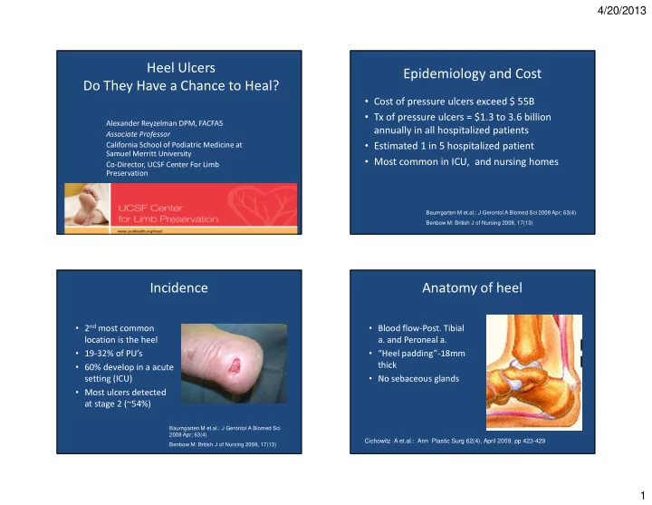

4/20/2013 Heel Ulcers Epidemiology and Cost Do They Have a Chance to Heal? • Cost of pressure ulcers exceed $ 55B • Tx of pressure ulcers = $1.3 to 3.6 billion Alexander Reyzelman DPM, FACFAS annually in all hospitalized patients Associate Professor • Estimated 1 in 5 hospitalized patient California School of Podiatric Medicine at Samuel Merritt University • Most common in ICU, and nursing homes Co-Director, UCSF Center For Limb Preservation Baumgarten M et.al.: J Gerontol A Biomed Sci 2008 Apr; 63(4) Benbow M: British J of Nursing 2008, 17(13) Incidence Anatomy of heel • 2 nd most common • Blood flow-Post. Tibial location is the heel a. and Peroneal a. • 19-32% of PU’s • “Heel padding”-18mm • 60% develop in a acute thick • No sebaceous glands setting (ICU) • Most ulcers detected at stage 2 (~54%) Baumgarten M et.al.: J Gerontol A Biomed Sci 2008 Apr; 63(4) Cichowitz A et.al.: Ann Plastic Surg 62(4), April 2009, pp 423-429 Benbow M: British J of Nursing 2008, 17(13) 1
4/20/2013 Risks factors Pathophysiology • PAD • CVA Skin breakdown • Age • Neuropathy • Pressure • Hip fractures Deep tissue injury (DTI) • Friction • Low serum albumin* “Reperfusion • Shear • Low Braden score* hyperemia” • Immobility Tissue Hypoxia • Diabetes* • Edema Increase Pressure, Shear, and Friction *Walsh J: Poster Adv. Wound Care, April 2006 Classification Classification Stage Description Unstageable wound Deep tissue injury 1 Non-blanching erythema/purple hue of skin, changes in temperature and sensation 2 Partial thickness skin loss i.e. blister or shallow crater 3 Full thickness skin loss involving necrosis of subcutaneous tissue 4 Full thickness skin loss with extensive necrosis to tendon, muscle, bone, or joint *Unstageable Ulcer with eschar-wound base can’t be assess *DTI Purple non blanchable area of intact skin which demarcates between 24-48 hours due to deep tissue destruction. Adapted from National PU Advisory panel (NPUAP) 2007 2
4/20/2013 Two Types of Heel Ulcers Plantar vs. Non-Plantar Heel Ulcers • Plantar ulcers • Non-plantar ulcers – Not decubitus in etiology – Low pressure over long – Occur in period of time ambulatory/younger (decubitus) Plantar Non-plantar individuals ulcers – Bedbound/ older Posterior – Heel walkers lateral and patients – Frequently occurs after a medial ulcers – Typically have poor failed Achilles arterial perfusion. lengthening procedure – Typically have adequate arterial perfusion ( Posterior heel ulcer) Management Offloading • Offloading is a must- in ALL stages -Prevent drop foot • Blood Flow has to be assessed -Reduce heel pressure below • Stage 1-2 foam, hydrocolloid dressings 32mmHg • Stage 3-4-Know when to debride, -meticulous skin controversial care • Nutritional assessment • DM related to poor outcomes Langemo D: Advances in Skin & Wound Care 2008, Farid KJ: Ostomy Wound Manag 2007; 53(4) Heel Pressure Ulcers: Stand Guard 3
4/20/2013 Offloading Results Plantar heel ulcers • Meta-analysis • 1457 subjects/104 studies • Pressure relieving surfaces were associated with significantly lower incidence of heel ulcers when compared with standard mattress • Insufficient research to conclude heel protective devices prevent heel ulcers. Langemo D: Advances in Skin & Wound Care 2008 Main Reasons For Failure Adjunctive Therapy • Lack of Arterial Osteomyelitis Perfusion • NPWT* • Bioengineered tissue** • Can’t adequately offload 4
4/20/2013 When to debride? Yes No Surgical Approach • Is the patient able to ambulate or transfer? • Is there adequate arterial perfusion? – Revascularization if needed • Surgical debridement Corticeal erosion – In office/clinic vs Operating Room • Partial vs. Total Calcanectomy Corticeal erosion 5
4/20/2013 Literature Review Results • Systematic review of literature for partial and • 60% of patients had no complications • 85% maintained ambulatory status post operatively total calcanectomies. Reviewed 26 publications that met the following criteria • 83% returned to ambulation with the use of normal or custom shoes with or without custom orthotics • Inclusion Criteria: • Patients with DM had nearly 5 times greater risk of – Calcaneal osteomyelitis major lower extremity amputation compared to – Partial or total calcanectomy patients without DM. – Ambulatory pre-operatively – Follow up of at least 12 mos Schade V., JAPMA 2012 Schade V., JAPMA 2012 6
4/20/2013 Post-OP Management Conclusion: • Systematic approach to heel ulcers should include: – Ambulation assessment – Vascular assessment – Infection assessment • If conservative therapy fails, surgical approach is warranted in the appropriate patients • Partial and/or Total Calcanectomy is a viable alternative to BKA. Minor exostectomy Thank You! 7
4/20/2013 Results When to debride? • Randomized clinical trial (level 2 evidence) Yes No • 338 adults, 3 pressure -reduction devices • 12 heel ulcers developed – Bunny boot=3.9% – Egg crate=4.6% – Foot waffle=6.6% • No statistical significance Gilcreast DM et.al.: J Wound Ostomy Continence 2005; 32 When to debride? CS CS 3 months Initial presentation 3 weeks 4 months 8
4/20/2013 Partial calcanectomy Results • Review 50 cases • Review 9 feet (8 pts) • 2/9 procedures BKA • 52-83% failure rate • Ambulatory patients prior • To evaluate factors that to surgery remained affects healing ambulatory – MRSA Randall D et.al.: JAPMA 2005 July/Aug; 95(4) – PAD • 20 PC, 11 TC during 10 year – Albumin levels period – Ulcer stage • 18 DM pts-Primary healing only in 4 pts • 65% Overall failure in DM Cook J et.al.: JFAS 2007, 46(4) Crandall, Wagner: JBJS Am 1981, 63(1) 9
4/20/2013 HPI Admission • 45 y/o HF, DM2 x 15 years presents to ED c/o • Nausea, vomiting, fever and chills x 2 days • WBC 20.5 painful left heel ulcer x 2 weeks. Began as a blister 2 nd to shoe rub, that progressed to • A1C=12.2 ulceration. She received tx in Mexico • Vasc: palp pulses except L PT (edema) consisting of Cipro and local wound care. She • Neuro: decreased protective sensation was d/c from care in Mexico 3 days prior to ED visit. She noticed increased pain, swelling, redness and drainage. Clinical Picture Hospital course Day of Admission • Zosyn 4.5 q 8H • Evening of admission- I&D with removal of all necrotic tissue 10
4/20/2013 Clinical Picture Culture results Post op day 4 • Tissue from 1 st I&D – Staph aureus – Strep B – Viridans Clinical Picture Post- op day 5 11
4/20/2013 Post op day 10 Post Ostectomy Day 2 Wound vac placed immediately after 2nd I&D Post debridement day 1 12
4/20/2013 Case-BH Hosp course on readmission • 64 y/o F with heel • Debridement of necrotic tendon, application ulcer, LE bypass by of wound vac vascular surgeon • Plastics did fasciocutaneous flap from calf and • Stagnant for 2 2 STSG from thigh months • DM, HTN, CAD • Heavy smoker • Caregiver Case-BH Case-BH Application 1 week post-application 13
4/20/2013 Case-BH Case-BH 2 week post-application 4 week post-application 14
Recommend
More recommend