Navicular Syndrome/Heel Pain Navicular Syndrome/Heel Pain Clinical - PowerPoint PPT Presentation
Navicular Syndrome/Heel Pain Navicular Syndrome/Heel Pain Clinical signs: Forelimb lameness, intermittent, progressive and insidious onset, usually bilateral. Stumbling Pointing toes to relieve pressure on DDFT Packing shavings
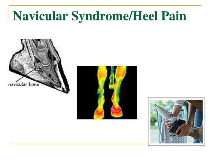
Navicular Syndrome/Heel Pain
Navicular Syndrome/Heel Pain Clinical signs: Forelimb lameness, intermittent, progressive and insidious onset, usually bilateral. Stumbling Pointing toes to relieve pressure on DDFT Packing shavings under front feet Decreased performance/stopping Short, stiff gait Chronic sequela-contracted heels, increased concavity of sole, toe bruising, may have enlarged digital vessels and increased digital pulses.
Anatomy of the Navicular Apparatus Associated structures: Navicular/distal sesamoid bone 30% of distal articular surface, acts as a fulcrum with DDFT Navicular bursa Deep digital flexor tendon (DDFT) Coffin joint Collateral (suspensory) ligament of navicular bone Distal sesamoidean impar ligament
The Equine Hoof Coffin joint Navicular bursa Impar ligament
Predisposing factors Conformation: Low/under run heels Small feet Long toes Improper shoeing Hard work on concussive surfaces Nutrition imbalances during younger years
Pathophysiology – why does it happen? 3 main theories - Contentious! Concussion leading to bursitis 1. Alters the flexor surface of the bone Villous hypertrophy, hyperplasia of synovial lining cells, venous congestion Remodeling of bone due to increased pressure of 2. deep digital flexor tendon (DDFT) Vascular 3. Arterial occlusion in foot, thrombosis and ischemic necrosis of navicular bone
Pathophysiology continued Syndrome most likely a mixed etiology: Age related changes-degenerative change in fibrocartilage on flexor surface of bone Adaptive remodeling of bone due to tendon stresses- cortical thickening Edema, congestion and fibrosis of marrow-cyst like- lesions Loss of proteoglycans in articular cartilage Bursitis Adhesions between DDFT and navicular bone
Differential diagnoses for navicular syndrome Puncture wounds to foot Fracture of navicular bone or distal phalanx Bruised sole Pedal osteitis Navicular bone Arthritis of coffin joint Corns Soft tissue injury such as lesions in the distal DDFT, impar ligament, or collateral ligaments.
Diagnosis Lameness exam Hoof testers-pain over center 1/3 of frog Distal limb flexion test/toe extension Palmar digital nerve block Intra-thecal analgesia of navicular bursa Coffin joint intra-articular analgesia
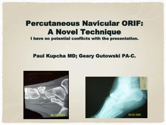
Diagnosis Radiographs 5 standard views most sensitive view 45 ° palmar proximal-palmarodistal oblique (navicular skyline view) Most significant changes likely to reflect navicular disease: Cyst like lesions in medulla Medullary sclerosis, and reduced cortico-medullary demarcation New bone growth/erosion on flexor surface Mineralisation in ligaments
Radiographs
Normal
Abnormal
Abnormal
Diagnosis continued… Nuclear scintigraphy Can see increased bone turnover MRI/CT More sensitive than radiographs Detects abnormal amount of fluid in coffin joint and navicular bursa Detects changes in bone such as fluid/edema Endoscopic evaluation of navicular bursa
Treatment options- no cure, only management Corrective trimming/shoeing Aim: balance the feet and straighten the pastern-hoof axis. Raise heels, shorten and roll toe to facilitate break-over Support across center 1/3 of frog Shoes: egg-bar, heart-bar, straight bar, natural balance
Treatment continued Drugs Analgesia 1. NSAIDS-phenylbutazone, naproxen, banamine, 1. aspirin Vascular modifying drugs 2. Isoxsuprine / Pentoxyfylline-vasodilators, alter 1. deformability of erythrocytes Dicumeral-blood thinner 2. Intra-thecal injection into navicular bursa 3. Polysulfated glycosaminoglycans-Adequan 4. Tildren-inhibits bone resorption 5.
Treatment continued Chemical “neurectomy”/cryoneurectomy -ablation of sensory fibers of palmar digital nerves Sarapin Clinical improvement lasts 2-3 months Unreliable
Treatment continued Palmar digital neurectomy Last choice option Careful selection of case, only if peri-neural analgesia of PD nerve eliminates lameness Successful in 65-70% of horses and lasts approximately 12-18 months Complications: Incomplete desensitization Regeneration of nerves Damage to DDFT Neuroma formation Change in hoof growth
Conclusion Multifactorial and controversial etiology No treatment is 100% successful Prognosis is always guarded: Many horses can continue to be useful if managed well, others may need to reduce level of work or even retire
Recommend


More recommend
Explore More Topics
Stay informed with curated content and fresh updates.