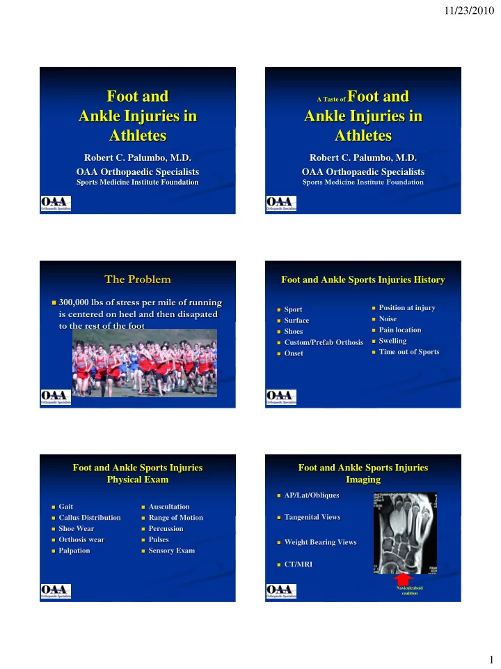

11/23/2010 Foot and A Taste of Foot and Ankle Injuries in Ankle Injuries in Athletes Athletes Robert C. Palumbo, M.D. Robert C. Palumbo, M.D. OAA Orthopaedic Specialists OAA Orthopaedic Specialists Sports Medicine Institute Foundation Sports Medicine Institute Foundation The Problem Foot and Ankle Sports Injuries History 300,000 lbs of stress per mile of running Position at injury Sport is centered on heel and then disapated Noise Surface to the rest of the foot Pain location Shoes Swelling Custom/Prefab Orthosis Time out of Sports Onset Foot and Ankle Sports Injuries Foot and Ankle Sports Injuries Physical Exam Imaging AP/Lat/Obliques Gait Auscultation Callus Distribution Range of Motion Tangenital Views Shoe Wear Percussion Orthosis wear Pulses Weight Bearing Views Palpation Sensory Exam CT/MRI Naviculcuboid coalition 1
11/23/2010 Foot and Ankle Sports Injuries Metatarsalgia Therapy Common overuse injury R.I.C.E. Early Gentle Motion described as pain in the Crushed ice best Whirlpool/Ultrasound forefoot that is associated Ice Massage Tilt Board with increased stress over Compression-Jones Strengthening the metatarsal head region Wrap Stretching!!! Crutches Often referred to as a symptom, rather than as a specific disease. Sesamoiditis Metatarsalgia SIGNS Common causes of Metatarsalgia Local Tenderness Sesamoiditis Interdigital neuroma (also known as Morton neuroma) Pain with Hyperextenion Avascular necrosis ( Frieberg’s Infarction) Rare Swelling Metatarsophalangeal Synovitis Inflammatory arthritis Synovitis/Inflammation from Repetitive Trauma Sesamoid Fracture Sesamoiditis Mechanism Incidence Stress Fracture Acute Fall from height (Ballet) Any age Tibial or Fibular Sesamoid Hyperext. Of MTP (football) Chronic-Stress Fracture(Runners) Osteochondritis Osteochondritis Female, 20’s lateral Sesamoid Kilman, F+A,3:220 1983 2
11/23/2010 Sesamoid Fracture Sesamoid Fracture X-RAY Acute Presentation AP/Lat/Oblique Reverse Oblique May mimic Turf Toe Tangential Views Bone Scan Treatment Depends on amount of Diastasis Sesamoid Fracture Sesamoid Fracture Acute Chronic Diastasis >2mm Treatment Bony Fixation U or J pad Soft tissue repair Firm Soled Shoes Diastasis < 2mm NWB 3 weeks if severe SLC 3-6 weeks Steel shank insole Prevent Hyperextension Sesamoid Fracture Sesamoid Fracture Surgical Treatment Excision of Fragment-Complications Displaced Fracture Migration of Hallux 10% Non-Disp Fx Not Resp to cast Immob. or shoe Persistent Pain 41-50% inserts x 12 wks Stiffness 33% Unrelieved Sesamoiditis/Bursitis Weakness 60% Osteomyelitis Richardson, F + A 7:29, 1987 Mann AOFAS 1985 3
11/23/2010 Sesamoid Fracture Turf Toe Late Repair Mechanism • Seventeen Patients Acute • Treated with Curretage and Bone Grafting Hyperextension of first MTP • Post-op SLC for Six Weeks Direct blow to heel with toe planted in dorsiflexion • Mean Follow-up 33 months Football Lineman • 15/17 Asymtomatic return to all Pre Injury Chronic Activities Repetitive valgus stress • 14/15 Healed by Tomography at 12 weeks Runner’s (Especially Cross-country) Anderson/McBryde AOFAS March 1991 Turf Toe Turf Toe Anatomy Treatment MTP Capsule No role for injections Articular Cartilage RICE, Shoe Mod. And Taping Great Toe Flexors If can’t jog w/in 3 wks. Sesamoids Consider Abductor Hallicus open treatment Late repair works Plantar Nerves Bones Coker, J Ark.Med Soc. 74:309 1978 Morton’s Neuroma Morton's Neuroma “Click" which is known as Mulder's sign There may be tenderness in the interspace Symptoms Rule out similar or concurrent problems Classically described as a burning pain in the forefoot Tenderness at one of the metatarsal bones can suggest can also be felt as an aching or shooting pain in the an overstress reaction (pre-stress fracture or stress forefoot fracture) in the bone. Pain may occur in the middle of a run or at the end of a An ultrasound scan can confirm the diagnosis and is a long run less expensive and at this time, at least as sensitive a test If your shoes are quite tight or the neuroma is very large, as an MRI the pain may be present even when walking An x-ray does not show neuromas, but can be useful to Occasionally a sensation of numbness is felt in addition "rule out" other causes of the pain. to the pain or even before the pain appears. 4
11/23/2010 Morton’s Neuroma Morton’s Neuroma Cause Contributing Factors An enlargement of the sheath Pronation of the foot can cause the metatarsal heads to of an intermetatarsal nerve in rotate slightly and pinch the nerve running between the the foot metatarsal heads Most Common – The third Chronic pinching can make the nerve sheath enlarge. intermetatarsal space As it enlarges it than becomes more squeezed and The second interspace increasingly troublesome. being the next most Tight shoes, shoes with little room for the forefoot, common location . pointy toeboxes can all make this problem more painful. Walking barefoot may also be painful, since the foot may be functioning in an over-pronated position. Morton’s Neuroma Morton’s Neuroma Orthotics – esp. for the Pronator Self-Treatment Injection of Steroid and Local Anesthetic Wear wide toe box shoes Occasionally injection of other substances to "ablate" the neuroma. Don't lace the forefoot part of your shoe too Surgical Removal of Neuroma tight Tips Make sure your feet are in supportive shoes that Wear shoes designed with a roomy toebox. do not squeeze your forefoot Wear shoes that have good forefoot cushioning. Use sport specific shoes. Fit your shoes with the socks that you plan to wear during aerobics activity. Frieberg’s Infarction Freiberg's Infraction Relatively uncommon AKA Avascular Necrosis, Osteonecrosis, Osteochondrosis General considerations Painful collapse of the head of the 2nd metatarsal Named “infraction” because it was originally thought May affect 3rd metatarsal head as well secondary to trauma Women to men by 5:1 Exact cause remains uncertain but thought to be one of Possibly because of shoes, i.e. stresses placed on the osteochondroses in adolescents toe by high-heeled shoes Osteochondroses are diseases that usually affect the epiphyses of growing bones resulting in necrosis most Length of second metatarsal thought to be a likely on a vascular basis, although the exact factor by some mechanism is not known Usually adolescents In others, Freiberg's may be due to a combination of Almost always unilateral trauma, and vascular insults 5
11/23/2010 Freiberg's Infarction Frieberg’s Infarction Imaging findings Clinical findings Early signs are sclerosis of 2nd Local pain, activity-related MT head and widening of joint space Tenderness Later there is fragmentation Stiffness and limp and collapse End result is flattening of head May produce “loose body ” Freiberg's Infarction Frieberg’s Infraction Treatment Surgical Complications Medical Premature closure of growth plate Immobilization and avoidance of weight- Loose bodies bearing to rest the joint Surgical Secondary osteoarthritis Various osteotomies, bone grafts, excision of the head, joint replacement have each been used alone or in combinations Midfoot Injuries Tarsometatarsal Sprains Considerable Disability Midfoot Dislocation Diagnosis Tarso-metatarsal Sprains Pain/Swelling over TMT Joint Metatarsal Fractures Flattening of Longitudinal Arch Metatarsal and Tarsal Stress Fractures Standing X-ray 6
11/23/2010 Base of 5th MT Fracture Tarsometatarsal Sprains Location Nondisplaced Tuberosity Avulsion Fracture Mechanism - Inversion Immobilize in NWB SLC for Eight weeks Heals Clinically-3 wks Orthotic Arch Support Radiograghically-6 wks Displaced Metaphyseal/Disphyseal ( Jones Fracture ) Reduction and internal fixation Mechanism – Supination Delayed union/non-union is common Jones Fracture Metatarsal Stress Fx Natural History SIGNS AND SYMPTOMS Fracture of Proximal diaphysis interrupts Recent Change in Distance intraosseous Blood Supply No relation to ht, wt., age Creates Zone of Relative Avascularity Tender on bone, not interspace Significant Delayed Union X-rays pos. at 3 -6 wks. Significant Refracture Bone scan is diagnostic Kavanaugh JBJS 60:776 1978 Smith F + A 13:143 1992 Jones Fracture Jones Fracture Treatment Presentation Type I Type I Acute Fracture, No IM Sclerosis NWB Cast Type II Type II Delayed Union, IM Sclerosis Non-Athlete treat Conservatively Type III Athlete treat w/ Curretage/Bone Grafting Non-union, Complete Obliteration of Type III Medullary Canal by Sclerotic Bone Curretage/Bone Grafting Torg JBJS 66:209 1984 Torg JBJS 66:209 1984 7
Recommend
More recommend