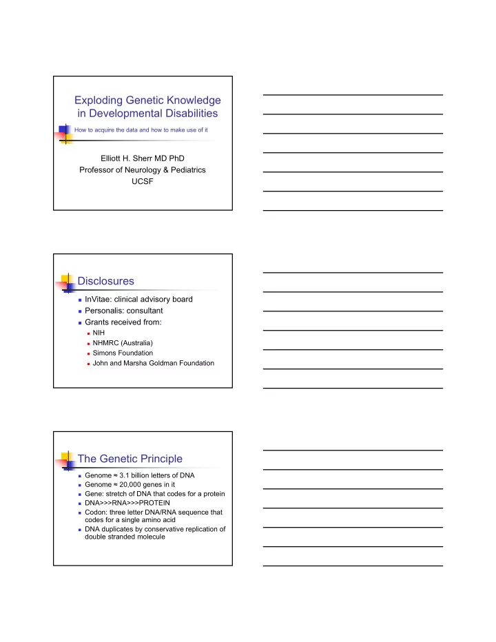

Exploding Genetic Knowledge in Developmental Disabilities How to acquire the data and how to make use of it Elliott H. Sherr MD PhD Professor of Neurology & Pediatrics UCSF Disclosures InVitae: clinical advisory board Personalis: consultant Grants received from: NIH NHMRC (Australia) Simons Foundation John and Marsha Goldman Foundation The Genetic Principle Genome ≈ 3.1 billion letters of DNA Genome ≈ 20,000 genes in it Gene: stretch of DNA that codes for a protein DNA>>>RNA>>>PROTEIN Codon: three letter DNA/RNA sequence that codes for a single amino acid DNA duplicates by conservative replication of double stranded molecule
The Genetic Principle The Genetic Code Autosomal Recessive
Autosomal Dominant: One gene A*A X chromosome linked inheritance A Aa Aa a AA Mutations: what are they? A genetic change that is deleterious Nonsense Missense Del/Dup Other (promoter, 3 ° structure, etc) Polymorphism A genetic change that may be positive or negative, but of less clinical impact Each of us has ≈ 3,000,000 polymorphisms and probably 10-20 “ silent ” mutations There is overlap between M & P
Nonsense Mutations Missense Mutations Polymorphisms If polymorphism is a single DNA letter; it is called a SNP (single nucleotide polymorphism) Some SNPs are common A = 80%; C = 20% Usually just two choices Can screen for common changes in large numbers throughout the genome easily Common differences each can contribute a small amount to common diseases Some SNPs are rare A = 99.9%; G = 0.1% As a collection these are important for disease Tools for assessing these are advancing rapidly
De Novo Genetic Mutations Mutation occurs just in that person Can occur during egg or sperm formation or just after fertilization Can be single base pair or chromosomal regions Occur more commonly in older parents Examples of de novo mutations Down syndrome (trisomy) Prader Willi syndrome (small chrom change) Dravet syndrome (point mutation) Chromosomal Ideograms SNP Micro ‐ Arrays Detect CNVs
Whole Exome sequencing gDNA Johnsen J M et al. Blood 2013;122:3268-3275 Genetics of Neurodevelopment Conditions & Identification Genetic Discovery; Mechanism • Down Syndrome (1866) • 1959 De novo • Neurofibromatosis (1882) • 1990 AD, De novo • Fragile X (1943 & 1969) • 1995 X ‐ linked • Prader Willi syndrome (1956) • 1981 De novo • Smith Lemli Opitz (1964) • 1998 AR • Angelman syndrome (1965) • 1987 De novo • Costello Syndrome (1977) • 2005 AD, De novo • Pitt ‐ Hopkins syndrome (1978) • 2007 De novo Expanding Number of Genetic Disorders
Expanding Diagnostic Yield in DD Genetics of Neurodevelopment: Advancing from research to the clinic Our patient C.O. in 2004
Our Patient in 2004 • Born Term, normal BW, Height and OFC • Developed GTC Sz at 9 months • At 1 yr had roseola infection and development was reported to decline after this • 19 months: bilateral cataracts and optic atrophy noted on eye exam • MRI at 23 months: hypoplasia of inferior cerebellar vermis cerebral hemispheres • Follow up scan at 46 months showed worsening brain atrophy , and smaller optic nerves and chiasm • NCS: 23 months: sensory neuropathy with delayed conduction, amplitudes normal, EMG normal • Sural nerve biopsy: thinly myelinated and 30% loss of axons • Loss of milestones , with cessation of speech. • Phys Ex: mild dysmorphism: hypertrichosis, synophyrus, deep ‐ set eyes with epicanthal folds, flat philtrum. Muscles had a doughy texture. Neuro: end gaze nystagmus with titubation, limb hypertonia, reduced reflexes, but plantar response was extensor. Diagnostic Eval on Patient C.O. • Metabolic Testing—negative – VLCFA, Phytanic Acid, plasmalogens – Lactate, pyruvate – Plasma lysosomal enzymes – CK, CDG, NCL • Genetic Testing ‐‐ negative – MELAS, MERRF, NARP, SCA6,7,8; DRPLA, Friedreich ataxia – Karyotype, Subtel, Microarray • ECHO, skeletal survey • Pause: Focus on clinical management – Labeled as MSS—neuropathy variant Exome Sequencing for C.O. in 2014 • KIF1A – R216H, c.647 G>A – Zygosity: Het; inheritance: De Novo – Kinesin: motor protein that travels on microtubule “tracks” • NID1 – T408K, c.1223 C>A – Zygosity; Het; Inheritance: De Novo – Nidogen: binds extracellular matrix
Progressive Volume Loss In KIF1A de novo Dominant Mutation Carriers KIF1A: Sites of Mutations Kinesin moving a vesicle on MT
Gliding assay https://valelab.ucsf.edu/images/movies/mov ‐ invitmtglid.mov Functional Consequences: Kinesin Mutations WT V220I ‐ Polymorphism A255V – Recessive T99M – De novo E253K – De novo R216C – De novo
MT Speeds in WT and Mut KIF1A SNP AR Lab Questions: Autism & Neurodevelopment START HERE Summary
The Sherr Lab Team Brain.ucsf.edu Elliott.sherr@ucsf.edu 415 ‐ 502 ‐ 8039
Recommend
More recommend