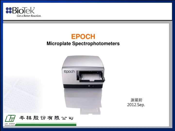

EPOCH Microplate Spectrophotometers 謝嚴蔚 2012.Sep.
BioTek’s product range: microplate instrumentation Washers Dispensers Pipetors Automation Absorbance Fluorescence Luminescence Multi-detection
Monochromator Optical System Data Xenon Flash Lamp Order Sorting Filters Diffraction Grating Optical fibers Photodiode Microplate Lens
Key Specifications – UV-Visble Spectrophotometer • Continuous spectrum – 200~999 nm Xenon Flash Lamp • Long life – 1 billion flashes • No warm-up required • No filters Monochromator Tg • Continuous wavelength selection Relative • Xe Spectral scans Energy Single channel optics Detector- Photodiode 100 250 1100 750 500 Wavelength 100 250 1100 750 500
Enhanced Reading Mode Ensures the highest quality readings ± 1% ± 0.005 ERROR OD Measured 0.923 OD Measured 1.234 OD Measured 2.700 NON ENHANCED OD Measured 0.675 OD Measured 2.678 ENHANCED WHY ? Plate 8 Flashes 8 Flashes 8 Flashes 8 Flashes 2 3 4 1 64 Flashes OPTICAL DENSITY If less than 2.0 OD then on to next sample If Greater than 2.0 OD then 64 more flashes, then on to next sample
Microplates (model dependent) � Microplates up to 20.3 mm in height – 6-well – 12-well – 24-well – 48-well – 96-well – 384-well � Terasaki trays – 60-well – 72-well – 96-well � Up to 8 Bio-Cells for 1 cm readings
Utilizes Bio-cell � 1.0 cm Quartz Cuvette for � Adapter Plate for Vertical Photometery Bio-Tek Microplate readers 1cm
Major Applications � Molecular Biology � Protein Quantitation � ELISA � Cell Viability/Cytotoxicity � Miscellaneous
Molecular Biology � Nucleic Acid Quantitation • DNA • RNA • Oligonucleotides � Nucleic Acid Quality Control • 260/280 Ratio (protein) • 260/240 (EDTA) • 260/270 (phenol) • 320 (particulates) ** UV Plate
Nucleic Acid Quantitation � Direct Quantitation : 280 nm (DNA Concentration = OD x 50) � Colorimetric Assays • Burton Method
Pathlength Correction • Beer-Lambert Equation: O.D. = ( ε ) x (Concentration) x (pathlength) Horizontal Photometry Vessel Absorbing solution ? cm Light Detector Source
Protein Quantitation 1. Direct quantitation: 280 nm 2. Colorimetric : – Lowry method: 660 nm – Bradford method: 595 nm – BCA method: 562 nm
ELISA 1 3 w w a a 1 s s h h Antibody Enzyme labelled antibody Sample antigen
ELISA � Horseradish peroxidase • OPD 450 nm & 492 nm • TMB 650 nm & 450 nm • ABTS 414 nm � Alkaline phosphatase • pnpp 405 nm • β -Galactosidase • ONPG 420 nm � Urease • urea bromocresol 588 nm
Cell-based assays Cell viability/cytotoxicity • MTT 570 nm • XTT 492 nm • WST-1 450 nm
Spectral scanning
Software Control � All control via USB port � Gen5 software – Operating system of choice – Included with instrument – Increased flexibility – Robotic applications
Any Questions?
Recommend
More recommend