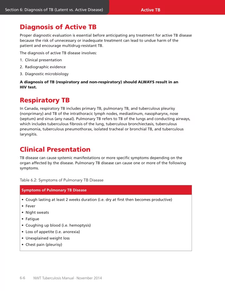

Active TB Section 6: Diagnosis of TB (Latent vs. Active Disease) Diagnosis of Active TB Proper diagnostic evaluation is essential before anticipating any treatment for active TB disease because the risk of unnecessary or inadequate treatment can lead to undue harm of the patient and encourage multidrug-resistant TB. The diagnosis of active TB disease involves: 1. Clinical presentation 2. Radiographic evidence 3. Diagnostic microbiology A diagnosis of TB (respiratory and non-respiratory) should ALWAYS result in an HIV test. Respiratory TB In Canada, respiratory TB includes primary TB, pulmonary TB, and tuberculous pleurisy (nonprimary) and TB of the intrathoracic lymph nodes, mediastinum, nasopharynx, nose (septum) and sinus (any nasal). Pulmonary TB refers to TB of the lungs and conducting airways, which includes tuberculous fjbrosis of the lung, tuberculous bronchiectasis, tuberculous pneumonia, tuberculous pneumothorax, isolated tracheal or bronchial TB, and tuberculous laryngitis. Clinical Presentation TB disease can cause systemic manifestations or more specifjc symptoms depending on the organ affected by the disease. Pulmonary TB disease can cause one or more of the following symptoms. Table 6.2: Symptoms of Pulmonary TB Disease Symptoms of Pulmonary TB Disease • Cough lasting at least 2 weeks duration (i.e. dry at fjrst then becomes productive) • Fever • Night sweats • Fatigue • Coughing up blood (i.e. hemoptysis) • Loss of appetite (i.e. anorexia) • Unexplained weight loss • Chest pain (pleurisy) 6-6 NWT Tuberculosis Manual - June 2014 NWT Tuberculosis Manual - November 2014
(Image from Medscape reference: Active TB Latent Section 6: Diagnosis of TB (Latent vs. Active Disease) Section 6: Diagnosis of TB (Latent vs. Active Disease) The physical exam is unlikely to fjnd any physical fjndings that are conclusive for a defjnite diagnosis of respiratory TB disease. This is the case even in advanced disease, since physical fjndings are not specifjc to just TB; however, it does give an overall impression of the status of the patient. Infected children under the age of 5 have a high risk of progression to severe forms of TB such as pulmonary TB, TB meningitis or miliary disease. Older children and adolescents present with symptoms of adult-type disease. Thus, they may present with the classic triad of symptoms: fever, weight loss and night sweats. Chest Radiography Chest radiography is a complimentary tool towards a diagnosis of pulmonary TB disease, and should be performed in any patient suspected of having pulmonary TB disease. It is important to be aware that chest radiography has substantial limitations in the diagnosis of pulmonary TB disease. Posterior-anterior [PA] and lateral radiographs are the standard views to see any chest abnormalities. Typical fjndings seen in individuals with a normal immune system, especially in reactivated TB, include: • Position – infjltrates in the apical-posterior segments of upper lobes or superior segment of lower lobes in 90% • Volume loss – this is a hallmark of TB disease because of its destructive and fjbrotic nature • Cavitation – this is seen at a later stage and depends upon a vigorous immune response, therefore often not seen in immunocompromised individuals Figure 6.4: Lung Cavitation in Left Upper Lobe http://img.medscape.com/pi/features/slideshow-slide/CXR/fjg11.jpg ) NWT Tuberculosis Manual - November 2014 NWT Tuberculosis Manual - June 2014 6-7
(Image from: CDC (2011), reference guide for physicians) Active TB Section 6: Diagnosis of TB (Latent vs. Active Disease) Figure 6.5: Chest Radiograph with Left Lower Lobe Cavity In children, PA and lateral radiographs are required to detect hilar and paratracheal lymphadenopathy, the most common features of pediatric TB. Parenchymal lesions may be found anywhere in primary disease and are typically, but not always, in the upper lobes. Cavitation is rare in children but can be seen in children with adult-type disease or caseating pneumonia. In patients who are immunocompromised (e.g. HIV infection, corticosteroid use, other), there are specifjc features found on CXR: • Hilar and mediastinal adenopathy • Non-cavitary infjltrates and lower lobe involvement Certain complications that may be diagnosed radiographically include: • Endobronchial spread of the disease (Tree in Bud appearance) • Pleural effusion • Pneumothorax 6-8 NWT Tuberculosis Manual - June 2014 NWT Tuberculosis Manual - November 2014
Recommend
More recommend