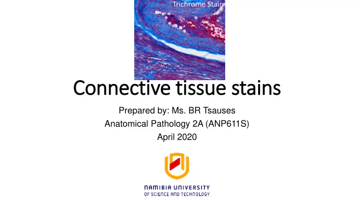

Connective tissue stains Prepared by: Ms. BR Tsauses Anatomical Pathology 2A (ANP611S) April 2020
Learning objective • Describe and apply appropriate staining techniques for connective tissue in histology.
Pre-learning Quiz • Please take the pre-learning quiz before proceeding with the presentation.
Trichrome tissues stains • Differential demonstration of the connective tissue • Term trichrome stain general name for a number of techniques to demonstration of: muscle, collagen fibers, fibrin and erythrocytes. • Masson's trichrome is a three-colour staining protocol used in histology. The recipes evolved from Claude L. • Pierre Masson's (1880 – 1959) original formulation have different specific applications, but all are suited for distinguishing cells from surrounding connective tissue.
Masson trichrome stain
Principle of Trichrome Stain • As the name implies, three dyes are employed selectively staining muscle, collagen fibers, fibrin, and erythrocytes. • The general rule in trichrome staining is that the less porous tissues are colored by the smallest dye molecule; whenever a dye of large molecular size is able to penetrate, it will always do so at the expense of the smaller molecule.
Masson trichrome (MT) stain principle • Others suggest that the tissue is stained first with the acid dye, Biebrich Scarlet, which binds with the acidophilic tissue components. • Then when treated with the phospho acids, the less permeable components retain the red, while the red is pulled out of the collagen. At the same time causing a link with the collagen to bind with the aniline blue.
Factors affecting trichrome staining • Tissue permeability and dye molecular size • Heat • Ph (should often be 1.5 - 4.0)
Effects of fixation • Routine fomaldehyde fixed tissues will produce optimal results. • Due to crosslink with protein leave few groups to react with trichrome dyes
Ideal fixative for MT • Bouins fixative • Zenker solution • Pecro-mercuric alcohols
Demonstration of elastic fibers • Verhoeffs van Gieson technique • Van Gieson Stain is used to differentiate between collagen and smooth muscle in tumors and to demonstrate the increase of collagen in diseases. • This method combines two or more anionic dyes and rely on differential binding by tissue components. • The differentiation is determined by a combination of differences in the relative size of the dye molecules, differences in the physical structure of the tissue, and differences in the amino acid composition of tissue Elements.
Principle of Van Gieson's Stain • Van Gieson's Stain is a mixture of Picric Acid and Acid Fuchsin. It is the simplest method of differential staining of Collagen and other Connective Tissue. • When using combined solutions of picric acid and acid fuchsin, the small molecules of picric acid penetrate all of the tissues rapidly, but are only firmly retained in the close textured, red blood cells and muscle. • The larger molecules of Ponceau S displace picric acid molecules from collagen fibres, which have larger pores, and allow the larger molecules to enter.
Ideal fixative • Buffered formaldehyde • Should contain potassium dichromate
Demonstration of van Gieson’s stain
Reticulin fibres • Reticulin fibres have little natural affinity for silver solutions so, they must be treated with potassium permanganate to produce sensitized sites on the fibres where silver deposition can be initiated. • The silver is in a form readily able to precipitate as metallic silver (diamine silver solution) • The Optimal pH for maximum uptake of silver ions is pH 9.0. A reducing agent, formalin, causes deposition of silver in the form of metal. Any excess silver in the unprecipitated state is removed by treating with hypo. Gold chloride treatment renders the preparation permanent and produces a neutral black colour of high intensity
Demonstration of reticular fibers
Alternative stain for reticular fibres • Gomori • Russell pentachrome stain
Self-assessment • Please complete the self-assessment activity provided.
References 1. John D. Bancroft, Christopher Layton and S.Kim Suvarna, (2013),Bancroft’s Theory and Practice of Histological Techniques,7thEdition, Elsevier, China 2. J.A.Kiernan,(2015)Histological and Histochemical Methods, 5thEdition, Scion, UK
Recommend
More recommend