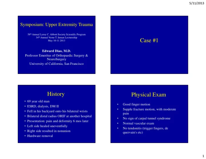

5/11/2013 Symposium: Upper Extremity Trauma 58 th Annual Leroy C. Abbott Society Scientific Program 34 th Annual Verne T. Inman Lectureship Case #1 May 10-11 2013 Edward Diao, M.D. Professor Emeritus of Orthopaedic Surgery & NeuroSurgery University of California, San Francisco History Physical Exam • 69 year old man Good finger motion • • ESRD, dialysis, DM II Supple fracture motion, with moderate • • Fell in his backyard onto his bilateral wrists pain • Bilateral distal radius ORIF at another hospital No sign of carpal tunnel syndrome • • Presentation: pain and deformity 6 mos later Normal vascular exam • • Left side healed uneventfully No tendonitis (trigger fingers, de • • Right side resulted in nonunion quervain's etc) • Hardware removal 1
5/11/2013 Wrist Deformity Initial Films Initial Closed Treatment ORIF: Hardware Failure 2
5/11/2013 Extreme Loss of Reduction Hardware Removed Distal Radius Nonunion Distal Radius Nonunion • Largest reported series of distal radius nonunion: • Traditionally, nonunion has been a rare complication of treatment of distal radius fractures Diego Fernandez, series of 23 patients. • Reported incidence 0.02% in reviews of thousands of • These authors advocated attempt at ORIF even cases, historically 1 • Recent trend toward open treatment may be when distal fragment was “small” increasing the incidence • This experience mostly predates modern internal • Most literature pre-dates modern methods of internal fixation fixation devices. 1 Bacorn R.W., Kurtzke J.F. A study of two thousand cases from the New Prommersberger KJ, Fernandez DL. CORR 2004 Feb;(419):51-6. • Nonunion of distal radius fractures. Clinic of Hand Surgery, Rhön-Klinikum, Bad • York State Workmens Compensation Board. J Bone Joint Surg. 1953; 38A, Neustadt, Germany. 643-58. 3
5/11/2013 Plan CT Scan To repeat the same thing and expect a • different result would be unwise Would need to address ulnar positivity • Articular surface is spared, so revision • open reduction and internal fixation might be rewarding, with ICBG Ulnar shortening performed at the same • time as revision ORIF Initial Post-Op 2 year follow up Functional • Painless • Stable wrist • Flex/Ext Arc 120deg • Pron 75 deg • Sup 70 deg • DRUJ stable • Right side has better • Motion than left • 4
5/11/2013 Case Presentation • 37-year-old woman s/p R elbow fracture 24 years ago. Case #2 • Status-post excision of radial head – Subsequent arthrosis of radial head – Proximal migration of the radius • DRUJ dysfunction and ulnocarpal abutment • Loss of supination Case Presentation • C/O Chronic right elbow pain. • C/O Chronic wrist pain as well • Limited ROM: – Good pronation to 90 degrees – But, supination approximately 60 degrees. • Tenderness over the ulnar side of the wrist when the wrist was in full extension and ulnar deviation. 5
5/11/2013 Diagnosis?:Essex-Lopresti Treatment?: Valgus stability • Ulna Shortening • Radial Lengthening • One Bone Forearm • Other 6
5/11/2013 Proximal Migration • The principal deformity after proximal translation is at the wrist: – The distal ulna sits dorsal and distal to the carpus, blocking supination and extension of the wrist. – Essex-Lopresti P: Fractures of the radial head with distal radio-ulnar dislocation: Report of 2 cases. J Bone Joint Surg Br 1951;33:244-247. • “the optimal solution to acute forearm dissociation would be internal fixation of the radial head.” 7
5/11/2013 Details of Radiocapitellar Joint Post-Op Xrays 8
5/11/2013 Final Result • Elbow room: 0/140 0 • Pronation: 90 0 Case #3 • Supination: 90 0 • Pain is significantly diminished DOS: G.B. 02/11/2009 •81 yo male, elite golfer •Severe OA on Right Elbow •Has a leg prosthesis •Golfs 18 holes daily 9
5/11/2013 DOS: Choices 01/14/2009 1. TEA 2. Fascial Interposition Arthroplasty 3. Fusion 4. ??? OR: 2/11/2009 • Scope R elbow • Complete loss of articular cartilage • Open Kocher approach, radial head excision • Push-pull test negative for radius migration • Annular ligament reconstruction 10
5/11/2013 G.B. – Post-Op F/U: 11/10/2010 (1 ½ yr P/O) • Recovery took 6 months • Plays 18 holes every other day • No swelling 11
5/11/2013 12
5/11/2013 FU: 1/11/12 (3 yrs P/O) V.B. - DOB:1962 • College basketball player – Forward • Right wrist scaphoid injury 30 years ago Case #4 • Now a 51 y.o. recreational athlete, still 6’8” and FIT • He can’t shoot the basketball without pain • He is having increasing trouble as an adult…can’t shoot the basketball anymore 13
5/11/2013 V.B. OR 9/14/2009 Diagnosis?: SNAC Wrist • Arthroscopic synovectomy, TFCC debridement Treatment?: • Proximal row radiolunate and radioscaphoid joints • Wrist Fusion preserved • Scaphoid partial excision distal radial portion (gross • Scaphoid Excision/Four degeneration) Corner Fusion • Radial styloidectomy • Scapholunate ligament degeneration noted • Anything more conservative? • Chronic scaphoid nounion pseudoarthrosis noted Post-op 9.23.09 V.B. Follow-up 5 ½ yrs 14
5/11/2013 V.B. Follow-up 5 ½ yrs OV 3.12.13 • No symptoms…I saw him when he brought his son in • Playing basketball, doing push ups, no pain • ROM is good 80% of normal • Fluoroscans did not show progression of disease C.G. 32 yo male, R hand dominant. • Case #5 R chronic scaphoid non-union. • OR #1: Volar approach + bone graft in distant past, • OR #2: 12/16/2009 – ORIF dorsal approach screw. • OR #3: 5/17/2010 – Revision with screw removal + • Bone graft substitute 15
5/11/2013 OV: 5/17/2010 OV: 5/6/2010 More Deformity! Post ORIF OR #2 – screw backed out OV: 10/18/2010 Post OR #3 Judgement Call • Screw S/P 2 operations with conventional • Screw plus bone graft fixations with bone substitutes. • Vascularized bone graft +/- fixation • Salvage procedure ?Now what should be done? 16
5/11/2013 The arc of reach of various distal radius pedicled bone grafts 1,2 ICSRA Fourth ECA Vascular Anatomy of the dorsal distal radius A. Shin & A. Bishop; JAAOS 2002 A. Shin & A. Bishop; JAAOS 2002 Vascularized bone graft mobilization and insertion into scaphoid nonunion A. Vascularized bone graft B. Dashed lines = donor site. 1,2 ICSRA is incisions of the first and identified second extensor compartments A. Shin & A. Bishop; JAAOS 2002 A. Shin & A. Bishop; JAAOS 2002 17
5/11/2013 OR #4 (10/18/2010) PRE-OP #4: 10/18/2010 Post-Op OR #4: 2/1/11 Scaphoid Fx - Advancements 1, 2, IMA Vascularized Bone Graft • Better implants – cannulated compression headless screws • Better surgical techniques – dorsal and volar approaches • Local vascularized pedicled bone grafts for malunions and nonunions • Faster rehab, reduced immobilization, better results 18
5/11/2013 O.L. – OR #1 2.3.10 • Severe rheumatoid arthritis of R thumb • Complete synovectomy of T thumb IP Case #6 • Complete release of medial and ulnar collateral ligaments • Osteectome • Arthrodesis O.L. – 2.3.10 3.17.10 19
5/11/2013 O.L. – OR #2 6.29.11 Pre-Op 2.69.11 • Rheumatoid arthritis/CREST syndrome w/ MCP jt arthritis, deformity and contracture • Tenolysis of flexor tendons x2 • Volar plate release MCP joint • Collateral ligament release MCP joint • Intrinsic release of 3 rd finger • MCP joint arthroplasty with implant Intra-Op 6.29.11 20
5/11/2013 Post-op 2.12.13 O.L. – OR #3 2.22.13 • Right hand Rheumatoid arthritis and scleroderma with PIP 2 nd and 3 rd joint severe arthropathies status post prior reconstructive surgery • 2 nd PIP joint resection arthroplasty and implant arthroplasty • 3rd PIP joint resection arthroplasty and implant arthroplasty • Rebalancing of Boutonniere/swan neck deformity, 2 nd and 3 rd fingers • Reconstruction radial collateral ligament, 2 nd and 3 rd finger with local tissue Post-op 3.5.13 Post-op 4.9.13 21
5/11/2013 Post-op 5.9.13 Post-op 5.9.13 Thank you! 22
Recommend
More recommend