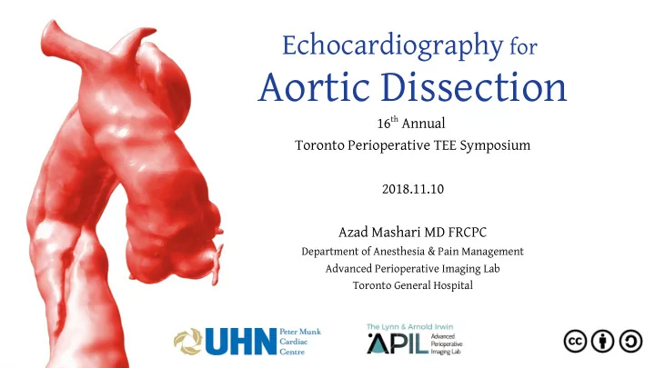

Echocardiography for Aortic Dissection 16 th Annual Toronto Perioperative TEE Symposium 2018.11.10 Azad Mashari MD FRCPC Department of Anesthesia & Pain Management Advanced Perioperative Imaging Lab Toronto General Hospital
This work is licensed under a Creative Commons Attribution 4.0 International License. For details see: https://creativecommons.org/licenses/by/4.0 You are fre ree to to: Share re — copy and redistribute the material in any medium or format Adapt — remix, transform, and build upon the material for any purpose, even commercially. The licensor cannot revoke these freedoms as long as you follow the license terms. Under r th the following te term rms: Attri ttributi tion — You must give appropriate credit, provide a link to the license, and indicate if changes were made. You may do so in any reasonable manner, but not in any way that suggests the licensor endorses you or your use. No additi tional re restri tricti tions — You may not apply legal terms or technological measures that legally restrict others from doing anything the license permits . Notices: You do not have to comply with the license for elements of the material in the public domain or where your use is permitted by an applicable exception or limitation. No warranties are given. The license may not give you all of the permissions necessary for your intended use. For example, other rights such as publicity, privacy, or moral rights may limit how you use the material.
Competing Interests No fjnancial disclosures :( Work supported by the Peter Munk Cardiac Center Foundation
Objectives At the completion of this presentation participants will be able to 1. Visualize & describe the anatomi mical cal relationships between thoracic aortic segments, tracheobronchial tree & esophagus to identify imaging windows and blind spots for TEE 2. Describe primary complications of acute TAD and corresponding clinica cal object bjectives es of f intraoper erative TEE during emergency repair surgery 3. Describe the ba basic c ec echoca cardiogr graphic c asses essmen ment of aortic dissection.
Intraoperative Echocardiography for Aortic Dissection Acute Type A Dissection for emergency repair Subacute Type A Iatrogenic Dissection Type A Dissection Traumatic Aortic Type B Dissection Dissection
Outline 1. Pathophysiology 2. Anatomy 3.TEE for emergency repair of ATAD ● Diagnosis ● Surgical planning ● Procedural guidance ● Post operative assessment
Pathophysiology of Aortic Diseases
Anatomy
https://i.pinimg.com/originals/d7/9e/2f/ d79e2f7c895d8ea328c9714acd3b4929.jpg
TEE TEE in Emergency Repair of Acute Type A Dissection “The primary purpose of intraoperative TEE is to detail ail the anat atomy my o of the dis dissectio ion an and t d to be better de defj fjne i its ph physio iolo logic gic co consequence ce” - Goldstein et al JASE 2015 Feb;28(2):119–82
Goals of TEE in Emergency Repair of ATAD 1. . Dia Diagnosis sis: Defjne anatomy & physiologic consequences of ATAD 2. . Procedura ral l pl plannin ing: Provide information relevant to key surgical decisions 3. . Mo Monit itori ring & gui uidance 4. . Post st-ope perative ive asse ssessm ssment
Goals: Diagnosis ● Assess presence of pe peric ricardial o dial or ple r pleura ural e efg fgus usio ion suggestive of aortic rupture ● Identify location of in intimal t imal tears ● Identify false & & t true rue lume lumens ● Defjne extent of dissection ● Asses ao aortic ic in insuffj ffjcie ciency ● Assess ventricul ricular r fun unctio ion ● Assess perf rfus usio ion of branching vessels
Goals: Diagnosis ● Assess presence of pe peric ricardial o dial or ple r pleura ural efg fgus usio ion suggestive of aortic rupture ● Identify location of in intimal t imal tears ● Identify false & & t true rue lume lumens ~30% ● Defjne extent of dissection 70% 70% ● Asses ao aortic ic in insuffj ffjcie ciency ● Assess ventricul ricular r fun unctio ion ● Assess perf rfus usio ion of branching vessels
Goals: Diagnosis – Luminal Truth Evangelista et al. Echocardiography in aortic diseases. Eur J Echocardiography. 2010 Sep;11(8):645–58
Question In what situation does the intimal fmap move towards ds the t true rue l lum umen in in systole? Which other typical fjndings of TL vs FL do not apply in this situation?
Goals: Diagnosis ● Assess presence of pe peric ricard ardial l or p or pleural ral e efg fgusion on suggestive of aortic rupture ● Identify location of inti timal mal te tears ars ● Identify false & & tru rue lu lume mens ● Defjne extent of dissection ● Asses aort ortic i insuffjc uffjciency ● Assess ve ventri ricular f ar fun uncti tion on ● Assess pe perf rfus usio ion of branching vessels
Goals: Diagnosis – Aortic Insufficiency A: Tear dilates Ao root & annulus – failure of coaptation B: : Asymmetric dissection depressed one leafmet below coaptation line C: Annular support disrupted, resulting in fmail leafmet D: Prolapse of intimal fmap through aortic valve in diastole, preventing coaptation Yas asmin S. Ham amiran ani et al al. Circulat ation. 2012;126:1121-1126
Goals: Diagnosis – Ventricular Function Generalized dysfunction associated with Acute AI Regional dysfunction associated Coronary artery injury/obstruction Coronary involvement: R > L
Right Coronary Artery
Right Coronary Artery
Left Main Coronary Artery
Left Main Coronary Artery
Goals: Diagnosis – Perfusion of Branches Arch & Visceral vessels ● Dynamic obstruction: Compression of TL by FL ● Static obstruction: Extension of dissection into or avulsion of branch
Caused by interposition of air-fjlled structures (tracheobronchial tree, lung) Often includes brac acheocephal alic & L common caro aroti tid Very rare for dissections to start or be limited to this area Dealing with the blindspot ● TTE suprasternal notch view ● Epiaortic imaging ● Bronchial balloon (“A-view” catheter)
Left subclavian artery http://pie.med.utoronto.ca/TEE/
Left common carotid http://pie.med.utoronto.ca/TEE/
Inominate artery http://pie.med.utoronto.ca/TEE/
Supresternal Notch View (TTE)
Goals: Diagnosis ● Assess presence of pe peric ricardial o dial or ple r pleura ural e efg fgus usio ion suggestive of aortic rupture ● Identify location of in intimal t imal tears ● Identify false & & t true rue lume lumens ● Defjne extent of dissection ● Asses ao aortic ic in insuffj ffjcie ciency ● Assess ventricul ricular r fun unctio ion ● Assess perf rfus usio ion of branching vessels
Goals: Procedur ural l Plann lanning ing Assist with Key Surgical Decisions ● Cannulation: – Venous: Central or femoral? – Arterial: Axillary or femoral? ● Arch repair? ● Aortic root repair/replacement? ● Aortic valve? ● Coronary bypass? ● Should pathology in descending aorta be addressed acutely?
Goals: Monit nitoring ring & & Pr Proce cedur ural al Guid uidance ance Dynamic process: extent & physiologic consequences can evolve Femoral cannulation: confjrmation of wire and cannula position Retrograde cardioplegia cannula EVAR guidance ● TEE can distinguish false &true lumens ● Avoid protruding plaques in landing zone
Two stage femoral venous cannula placement: guidewire
Two stage femoral venous cannula placement: guidewire
Two stage femoral venous cannula placement
Two stage femoral venous cannula placement
Goals: Post Operative Assessment ● Confjrm exclusion of entry tear and any proximal ● Ventricular function ● Aortic valve function ● Adequacy of fmow in descending thoracic aorta
References ● Goldstein et al. Mu Multi timod modality I ty Ima maging o of Di Disea seases o es of the the T Thoracic Ao Aorta ta i in Ad Adults. JASE 2015 Feb;28(2):119–82. ● Evangelista et al. Echo hocard rdiograph phy y in a n aorti tic d disea sease ses. Eur J Echocardiography. 2010 Sep;11(8):645–58. ● Erbel R et al. 2014 ESC G Guidel elines nes o on the the di diagnosi sis s and nd trea treatmen ment o t of a aorti tic disea seases es: Document covering acute and chronic aortic diseases of the thoracic and abdominal aorta of the adult. Eur Heart J. 2014 Nov 1;35(41):2873–926. ● David TE. Surg rgery f y for or a acute t type A a e A aor orti tic d disse ssecti tion. J Thorac Cardiovasc Surg. 2015 Aug;150(2):279–83.
APIL.ca azad.mashari@uhn.ca
Acknowledgements Jo Carroll, Sarah Russell & the Organizing Team ● Max Meineri, Joshua Hiansen, Jacobo Moreno, Annette Vegas, Jackie Cade, Patricia ● Murphy & the PMCC Foundation UHN Department of Anesthesia & Pain Management ● Anesthesia Associates ● Thank you!
Recommend
More recommend