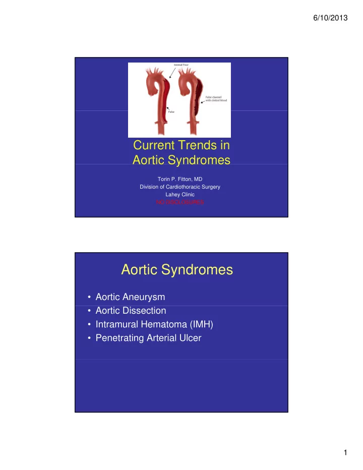

6/10/2013 Current Trends in Aortic Syndromes Aortic Syndromes Torin P. Fitton, MD Division of Cardiothoracic Surgery Lahey Clinic NO DISCLOSURES Aortic Syndromes • Aortic Aneurysm • Aortic Dissection • Intramural Hematoma (IMH) • Penetrating Arterial Ulcer 1
6/10/2013 Objectives • Spectrum of Aortic Syndromes • Historical Milestones Hi t i l Mil t • Risk Factors & Epidemiology • Aortic Imaging Modalities • Classification Schema • Operative Techniques & Outcomes • Operative Techniques & Outcomes • Endovascular Repair [TEVAR] Historical Milestones • 1760 Nicholls autopsy of King George II reveals intimal tear & aortic wall hematoma after he collapsed while straining on a commode 1761 Morgagni coins term ‘ aortic dissection ’ • 1761 Morgagni coins term aortic dissection • 1930 Erdheim describes histologic changes- cystic medionecrosis • 1935 Gurin attempts repair by fenestrating iliac artery • 1948 Contrast angiography introduced for diagnosis • 1955 DeBakey surgically treats patients with primary repair • 1956 Cooley & DeBakey: Cardiopulmonary bypass with selective anterior cerebral perfusion used (mainstay of contemporary aortic surgery) g y) • 1965 DeBakey classification schema; • 1965 Wheat medical treatment to decrease bp & wall stress (dp/dt) • 1975 Griepp uses hypothermic circulatory arrest • 1990 Endovascular stents 2
6/10/2013 There is no disease more conducive to conducive to clinical humility than aneurysm of the aorta . Sir William Osler Risk Factors for Aortic Syndromes • Connective Tissue Disorders – Marfan Syndrome – Ehlers-Danlos Syndrome – Loeys-Dietz Syndrome Loeys Dietz Syndrome • Degenerative – Atherosclerosis/Hypertensive – Cystic Medial Necrosis • Congenital – Bicupsid Aortic Valve/Ascending Aortopathy – Coarctation of the Aorta • Inflammatory • Inflammatory – Arteritis/Vasculitis – Bacterial/Syphylitic • Familial predisposition Aneurysm/Dissection • Aortic Dissection 3
6/10/2013 Epidemiology of Thoracic Aortic Aneurysms • 30-60,000 deaths/year • 18 th most common cause of th death (more than HIV) • Frequency increasing • Circadian-Diurnal variation • Exact prevalence unknown • Anterior MI • Sudden cardiac death Epidemiology of Aortic Dissections • Estimated 2.6-3.5/100,000 patient years • IRAD Database: • Mean age 63 • 2:1 male predominance • Older patients: 72% have HTN, atherosclerosis, or previous heart surgery previous heart surgery • Younger patients have Marfan’s or bicupsid AoV Tsai et al, Circulation 2005 4
6/10/2013 Criteria for Intervention on the Diseased Aorta Diagnosis of Aortic Syndromes • Poor prognosis after dissection/rupture mandates intervention before it occurs • Biomarker identifying risk/presence of aortic syndromes would help differentiate chest pain syndromes with would help differentiate chest pain syndromes with different management • Matrix metallo-proteinases • Circulating smooth muscle myosin chain • Inflammatory markers: CRP, fibrinogen, elastin fragments fragments • Ribonucleic acid signatures • Currently no available, reliable biomarker assay Elefteriades et al, J Am Coll Cardiol 2010 5
6/10/2013 Annual Rates of Complications Related to Aortic Size Elefteriades et al, J Am Coll Cardiol 2010 Hinge Points Defining Lifetime Risks Size best criteria determining intervention Imaging most reliable diagnostic tool Elefteriades et al, J Am Coll Cardiol 2010 6
6/10/2013 Aortic Imaging Modalities Goals of Aortic Imaging • Confirmation of Diagnosis Confirmation of Diagnosis • Classification • Tear localization & extent (dissection) • Indicators of Emergency • Pericardial/Mediastinal/ Pleural hemorrhage g • Arch & Side-branch involvement Aortic Imaging Modalities • Each imaging modality is accurate for a specific portion t f ifi ti of aorta • Need multiple modalities • Compare images versus all previous images • Changes < 3 mm imperceptible • Changes < 3 mm imperceptible because aorta is dynamic structure 7
6/10/2013 Echocardiography • Available • Crisp images • Aortic root to STJ • Assess for AI, tamponade, LV fxn • TEE better for arch, prox desc Ao Computed Tomography Widely available Excellent for distal ascending aorta, arch & head vessels, descending thoracic & abdominal aorta Axial cuts at root/valve level make measurement difficult 8
6/10/2013 Computed Tomography/M2S CT Axial Coronal & Saggital CT Axial, Coronal & Saggital cuts facilitate 3-D reconstruction using specialized operative planning software MRI • Beautiful images • Highly accurate • Limited availability, especially in emergencies 9
6/10/2013 Indications for Operative Intervention Operative Indications For Ascending & Arch Aorta • Size >5.5 cm atherosclerotic, degenerative or hypertensive aneurysms hypertensive aneurysms • Size >5.0 cm with bicupsid aortic valve, connective tissue disorder, familial history of aneurysms/dissection • Enlargement >0.5cm/6-12 months • Symptomatic Aortic Valve Regurgitation S t ti A ti V l R it ti 10
6/10/2013 Operative Indications for Descending Aorta Size greater than 6.0 cm • >5.5 cm connective tissue disorders, familial >5 5 ti ti di d f ili l • history aneurysm/dissection Median size 5.4 cm in Type B dissection as indication – is usually aneursymal enlargment of false lumen Enlargement > 0.5 cm/6-12 months • S Symptomatic aneurysm t ti • – Persistent pain, rupture, visceral malperfusion, Operative Indications for Aortic Dissections Tsai et al, Circulation 2005 11
6/10/2013 Aortic Syndrome y Classification Schema Aortic Dissection Classification • Classification – Multiple systems p y – All based on location of intimal tear – DeBakey & Stanford classifications used most frequently – Stanford A: • Any dissection involving ascending aorta no matter ascending aorta no matter primary tear – Stanford B: • Dissection involves only the descending aorta 12
6/10/2013 Crawford Classification Extent Origin and Location Distal to L SCA to above renal I arteries Distal to L SCA to below renal II arteries 6th IC space to below renal arteries III 12th IC space to iliac bifurcation 12th IC space to iliac bifurcation IV Below 6th IC space to above the V renal arteries Penetrating Arterial Ulcers Intramural Hematoma Advanced imaging g g has defined precursors to aortic dissection/rupture Likely all part of a continuum Tsai et al, Circulation 2005 13
6/10/2013 Intramural Hematoma • Collection of blood within the wall of the aorta without an identified of the aorta without an identified intimal tear • Proposed pathology: – Vasovasorum rupture/aortic media abnormality – Continuum of aortic dissection: noncommunicating aortic dissection with thrombosed false lumen Penetrating Arterial Ulcer • Deep ulceration of atherosclerotic plaques can lead to: – IMH – Aortic Dissection – Perforation – Pseudoaneurysm • Treatment of IMH/PAU based on aortic dissection classification 14
6/10/2013 Operative Techniques p q and Outcomes Pioneers of Aneurysm & Dissection Surgery Operative Mortality 60% 60% Cooley DA, DeBakey ME JAMA 1956; 162:1158 15
6/10/2013 Era of Era of Modern Modern Aneurysm Surgery Aneurysm Surgery Bentall-Bono Procedure Composite AVR • Aortic Valve • Coronary button (Bono modification) • Asc Ao Graft 16
6/10/2013 2 Pages 3 Fig 1 Ref Cabrol Modification 17
6/10/2013 Ascending Aorta and Hemiarch Replacement Total Arch and Elephant Trunk Replacement 18
6/10/2013 Valve-sparing Aortic Root Replacement Remodeling (Yacoub) Reimplantation (David) Repair of Type B Dissection Thoracoabdominal Aneurysm 19
6/10/2013 Outcomes of Aortic Dissection • Operative mortality – Type A: 7 -12% – Type B: 35-75% • Surgical Long-term Survival – 1, 5, 10, 15 year • Type A: 67%, 55%, 37%, & 24% • Type B: 56%, 48%, 29%, & 11% • M di Medical Long-term Survival l L t S i l • Type B: 73%, 58% – no significant survival difference versus surgical therapy Tsai et al, Circulation 2005 T horacic E ndo- V ascular A neurysm R epair p 20
6/10/2013 Endovascular Repair • Appealing alternative option given open repair mortality 35-75% • Many of those dying in medical therapy arm did die from complications of the dissection • Goals of Endovascular Repair • Reconstruction of segment containing entry tear • Induction of thrombosis of false lumen • Reestablishment of true lumen and side-branch flow Endovascular Devices MEDTRONIC TALENT Stanford Device GORE TAG COOK TX2 21
Recommend
More recommend